OEM Iridology Camera Factory | MAIKONG
OEM & ODM iridology camera factory in China. Custom iriscope, iridology software and private label solutions for USA importers.

How to design a clear, clinic‑ready iridology report
November 1, 2026
Quick answer
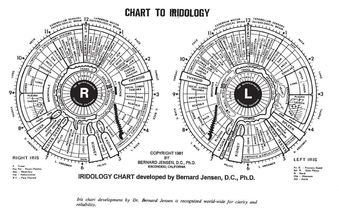
Design a clinic‑ready iridology report by (1) stating its purpose and limits up front, (2) including a concise client summary and consent record, (3) providing high‑quality annotated iris images with orientation markers and a mapped iridology chart, and (4) exporting standard, editable files so the record can be reviewed and compared later. Use controlled capture (consistent illumination, focus, white balance) and software that preserves original files plus an annotated copy. Follow the checklist and layout below to make reports clear to clients and clinically useful to practitioners.
Contents
What is iridology — one concise definition for Canadian practitioners
Iridology is a complementary observational system that records iris and periorbital features to support practitioner assessment and client education. It is a visual, non‑invasive method for documenting iris signs and changes over time; it is not a medical diagnostic test. We present report design advice here with that boundary in mind.
Report purpose and realistic limits: what a report should and should not claim
A clear iridology report has two practical goals: to communicate findings to the client in plain language, and to provide a reproducible record for the practitioner’s clinical notes. It should:
- Describe observed iris features and their mapped location on a standard chart.
- State practitioner interpretation, recommended follow‑up or lifestyle suggestions where appropriate.
- Include the capture metadata (camera model, lens, capture settings, date/time) so future comparisons are meaningful.
Do not use the report to claim medical diagnoses, prognoses, or to replace laboratory or physician assessment. Keep clinical language proportional to the evidence and always document that iridology observations are part of complementary practice.
Minimum delivery your client expects: the essential report elements
Clients and clinics expect a report that they can read and use later. At minimum include:
- Front page: client name, date of birth or client ID, photo date, practitioner name and clinic contact.
- Consent and intake summary: written consent for iris photography, brief medical history relevant to the consultation, and signature or e‑consent record.
- Client summary: plain‑language overview of observations and any practitioner recommendations or notes for follow‑up.
- High‑resolution annotated iris images (left and right) with orientation markers and scaling where possible.
- Iridology chart mapping showing zones used in your system and links to the annotated images.
- Practitioner notes and references to any client resources or next steps.
Image evidence: capture, labelling and quality checklist (use with MAIKONG camera specs)
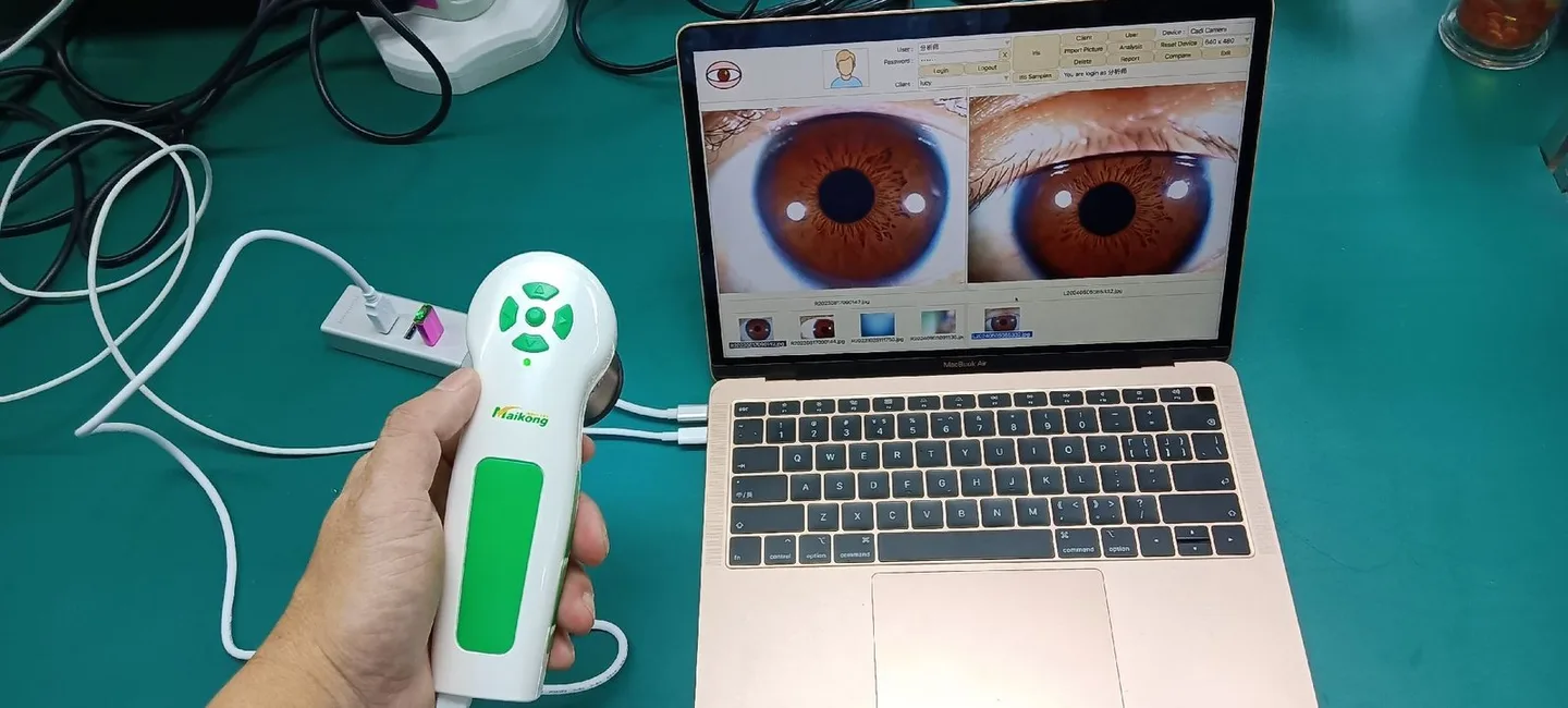
Image quality is the single most common cause of rework. Control these capture variables every time:
- Framing and orientation — always include a small periorbital area, show the nasal side (mark left/right), and keep the eye centred with consistent distance.
Checklist for an acceptable capture before leaving the client:
- Iris in focus with visible crypts/pigment patterns.
- No specular highlights across the iris disc; reflections controlled.
- Correct left/right label and orientation marker present.
- Skin colour not oversaturated; white balance OK on preview.
- Original (raw or lossless) file saved plus an annotated copy exported.
Suggested report layout: front page, client summary, annotated iris images, chart mapping, practitioner notes

Keep most reports to 2–6 pages so clients read them and clinicians can file them easily. A pragmatic layout:
- Page 1 — Cover & consent snapshot: client ID, session date, signed consent excerpt, reason for visit.
- Page 2 — Executive summary: three‑line plain‑language observation and suggested next steps.
- Pages 3–4 — Annotated images: left and right iris photos at the largest practical size with numbered markers and short captions.
- Page 5 — Chart mapping: iridology chart showing zone labels and arrows referencing the photo markers.
- Final page — Practitioner notes & metadata: capture device (make/model), software version, capture settings, and appointment notes.
Include a small legend explaining your chart terms and symbols; this reduces misinterpretation by clients and other practitioners.
How to annotate iris images practically (markers, zones, orientation and consistent terminology)
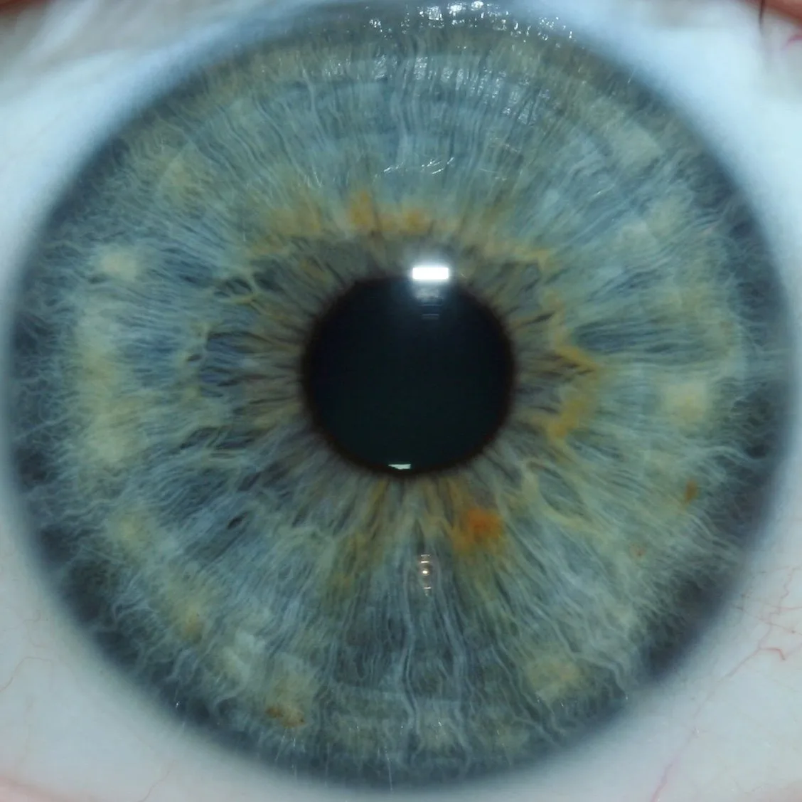
Annotations should be minimal, consistent and reproducible:
- Numbered markers (1, 2, 3…) rather than long labels on the photo; provide a keyed legend beneath the image.
- Use colour coding sparingly (e.g., red = focal area, blue = structural pigment change) and keep the legend fixed across reports.
- Always show the nasal‑temporal orientation (N/T) and which eye (L/R). A small compass arrow is better than text-only labels.
- Map markers to your iridology chart with zone numbers to make later comparisons precise.
- When noting minor changes, include relative language (“pronounced”, “mild”) and, when available, an earlier image comparison annotated side‑by‑side.
Choosing resolution and when 5MP vs 12MP matters for reporting

Higher pixel count gives more cropping latitude and finer visible detail, but it also increases file size and processing needs. Use higher resolution when you intend to zoom into small iris details or to print large images; lower resolutions are adequate for screen reports and routine clinic records.
| Model | Sensor / resolution | Illumination | Software & OS |
|---|
Trade‑off guidance: pick 5MP for faster capture, lower storage and routine documentation; pick 12MP if teaching, publications, or high‑magnification comparison are regular needs. Confirm your clinic network storage and backup strategy before selecting a higher‑megapixel workflow.
Workflow steps to produce a usable report during a brief clinic consultation

Design the workflow so the capture and basic report drafting fit into a single consultation without rework. A practical sequence:
- Prepare client file and consent form before the session; confirm identity and the intended use of images.
- Capture both eyes with the same lighting and focus settings; check images against the quality checklist and re‑capture if required.
- Import originals into analysis software immediately; save originals to archive and create an annotated working copy.
- Place annotated photos into your report template, add the chart mapping and a short client summary written in plain language.
- Export a client PDF and save the master files (original + editable) into the clinic record system with a consistent filename and metadata (see next section).
Automate repetitive tasks where your software allows it (batch export, standard legends, template insertion) to reduce administrative load.
Record keeping and client consent: minimum administrative fields and file naming
Keep consistent records for legal clarity and clinical continuity. Minimum administrative fields to include in the client record are:
- Client full name, unique client ID, date of birth (or age), contact details (as per your local privacy rules).
- Capture date and time, practitioner name, and clinic location.
- Device used (camera model), software name and version, and capture settings you altered (if any).
- Signed consent for photography and data retention, with clear statement of intended uses.
Suggested filename convention for easy retrieval: ClinicID_ClientSurname_DOB_YYYYMMDD_Eye_L/R_original.ext (e.g., CLN23_Smith_19780912_20261025_R_original.tif). Keep an internal index or database linking file names to the client record.
Common mistakes that reduce report value and how to avoid them
- Poor lighting and reflections: Control lighting with a ring or LED array; confirm no bright hotspots on the iris before saving.
- Missing orientation labels: Always mark left/right and nasal/temporal; failure to do so makes longitudinal comparison unreliable.
- Sole reliance on annotated images without original files: Preserve originals; annotations can obscure raw evidence needed for later review.
- Inconsistent terminology: Agree clinic‑wide chart labels and abbreviations; include a legend on every report.
- Not recording device/software metadata: Save camera model and software version on the report to explain why images look different if software updates change processing.
Evidence and limitations — what MAIKONG product facts change your choices
We supply imaging hardware and software that can reduce rework when used correctly. Relevant supplied details to check when choosing equipment:
Limitations to be explicit about: manufacturer provided specifications are descriptive; they do not, on their own, establish clinical accuracy or regulatory clearance for medical diagnostics. Always align your reporting practice with local professional and legal obligations in Canada.
Practical next step
If you want a clinic template or need configuration advice for a MAIKONG setup (camera model, software language pack, or export options) email us with your clinic use case, preferred OS (Windows or macOS), intended archive format and the approximate number of monthly captures. Pricing and configuration advice are available on inquiry.
Request configuration advice or a quotation — lucy@myiridology.us
FAQ (short)
Which file format should I archive for legal records?
Archive a lossless original (TIFF if possible) and also keep an annotated PDF for client delivery. If storage is limited, use the highest‑quality JPEG but ensure you retain one lossless original elsewhere.
Do MAIKONG cameras work with our clinic Mac workstations?
Important Disclaimer
Important Disclaimer: Content provided here is for educational and informational purposes only and does not constitute medical advice, diagnosis, or treatment. Iridology is a complementary wellness practice and is not a substitute for medical examination, diagnosis, or treatment. Iridology cameras and iriscopes are professional wellness tools intended for trained users. Ensure use of equipment complies with applicable Canadian laws, regulations and professional requirements.
This content is provided for learning, information sharing and professional communication only. It is not medical advice and is not a basis for treatment or diagnosis. Confirm suitability, operation and purchasing requirements against the product manual, local regulations and qualified professional guidance.
Related resources
Related resources
MAIKONG IRIDOLOGY CAMERA
MAIKONG IRIDOLOGY SOFTWARE Installation and operation
Exhibition


Company Certificates


Contact US Online WHATSAPP:

Maybe you like also
Copy©2026-2027