
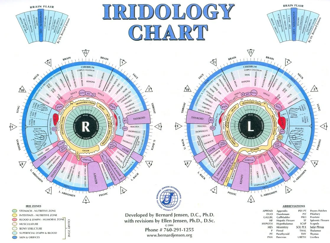
Iridology Chart Workflow for Canadian Centres
Read a comparable provider chart by documenting a high-quality bilateral iris image, aligning that image to the chart’s zone map, noting structural cues (fibre pattern, radial furrows, pigments, rings and focal spots), and recording repeatable observations in the client file. Before interpreting, ensure a trained practitioner reviews findings and that any health concerns are referred to regulated medical professionals. This iridology chart workflow emphasises image quality, consistent mapping, and careful wording to avoid medical claims.

Iridology Image Samples: Importer Checklist
Canadian importers should request representative iridology image samples including unedited TIFFs or high‑quality JPEGs for left/right iris, sclera, a magnified sector, capture settings and EXIF metadata, plus the vendor software export or PDF. Evaluate resolution, evenness of illumination, focus across the iris, LED illumination pairs for reflections or hot spots, and whether annotated exports map to the raw files before accepting shipment or listing a device. Specify model names (for example MAIKONG MK 980U or MK-9822U) and intended workflow to get verifiable, reproducible examples.