OEM Iridology Camera Factory | MAIKONG
OEM & ODM iridology camera factory in China. Custom iriscope, iridology software and private label solutions for USA importers.

Iridology examples: what iris image samples look like and what Canadian importers should check
September 18, 2026
Quick answer
comparable provider examples are the actual iris and sclera photographs, processed images and annotated charts suppliers provide to show camera performance and typical outputs. For Canadian importers the practical requirement is straightforward: request representative raw captures (left/right iris, sclera, close-up of pupil/limbus), the capture settings and an unedited TIFF or high-quality JPEG per sample, plus the software export used. Evaluate those files for resolution, evenness of illumination, focus across the iris, and embedded metadata before you accept a shipment or list a device. When you request samples, name the models you are considering (for example MK 980U or MK-9822U), the sample types you need, and the target workflow (clinic record, training, retail listing). This lets vendors deliver verifiable examples that answer your technical questions rather than promotional claims.
Contents
Direct answer: What “comparable provider examples” means for importers in Canada
We mean concrete image files and related capture data that prove a camera will meet your downstream needs: raw camera captures (left and right eye, sclera, magnified iris sectors), the capture settings (sensor size, resolution, illumination mode), and the software-exported reports or annotated charts. For procurement decisions you need files you can open and inspect locally, not only screenshots or marketing pictures.
Quick definition: comparable provider images and common image types you will see
In practice the phrase “comparable provider examples” covers a small set of standard image types. Expect to see:
- Full-frame iris photos — the whole iris centred, pupil visible, taken with standard iris lens.
- Sclera or conjunctiva shots — wider-angle images that include the white of the eye and surrounding tissue.
- Magnified sectors — close-ups of radial fibres, pigmentation, or spots for teaching or record comparison.
- Dual-light captures — the same eye with different LED combinations (e.g. iris-only vs. sclera illumination) to show contrast control.
- Exported analysis screenshots — software overlays or charts produced by comparable provider software (useful only as reference; they are not medical evidence).
Practical examples importers should recognise — what each image shows operationally
Each sample serves a purpose when you evaluate a shipment or supplier claim:
- Full-frame iris: confirms focus across the iris radius, chromatic balance and sensor resolving power. A blurred pupil edge or uneven radial detail suggests optics or focus issues.
- Sclera shot: demonstrates how well the device handles broader lighting and whether reflections or hot spots appear in peripheral regions.
- Magnified sector: shows resolving detail — whether the camera can capture micro-patterns without pixelation at your intended working distance.
- Dual-light pairs: let you assess whether the device’s LED configuration gives consistent contrast when toggling illumination modes (useful in MAIKONG models with multiple LED controls).
- Annotated export: valuable for product listings and training materials; verify that the software overlay matches the raw image rather than being generated from a different source file.
How example images are captured: basic camera, lighting and capture workflow for reliable samples
Request a short capture log with every sample. A minimal, practical capture workflow you should specify:
- Subject setup: neutral background, stable headrest or chinrest, natural sit height.
- Camera mode: single-shot capture with LED mode documented (which LEDs were on: iris/sclera combination).
- Lens and magnification: note if the 30X iris lens or standard lens was used.
- File output: uncompressed TIFF or high-quality JPEG (minimum preferred), with capture timestamp in metadata.
These elements let you reproduce the capture and validate whether the images represent routine clinical use or studio conditions.
Real-world use scenarios for example images in Canadian distribution and resale
For Canadian importers, image examples are used at three practical points:
- Technical acceptance: inspect samples on arrival to confirm resolution, illumination quality, and absence of manufacturing defects.
- Marketing and listings: images used in product pages, training handouts and trade-show displays need to be high-resolution and accurately labelled (model, capture settings).
- Training and customer support: teaching materials and demonstration files should include paired raw and annotated examples so your local trainers can teach capture technique consistently.
What good example image files include (format, resolution, metadata)
Ask suppliers to provide:
- Raw or minimally compressed image: TIFF preferred; high-quality JPEG acceptable for listings.
- Resolution and sensor notes: pixel count and stated maximum capture resolution (we will supply exact model specs on request).
- EXIF or sidecar metadata: capture timestamp, illumination mode, lens identification, software version used to save the file.
- At least one unedited left-eye and right-eye pair plus one magnified sector and one sclera shot per model for direct comparison.
Device examples and compatibility notes relevant to image examples
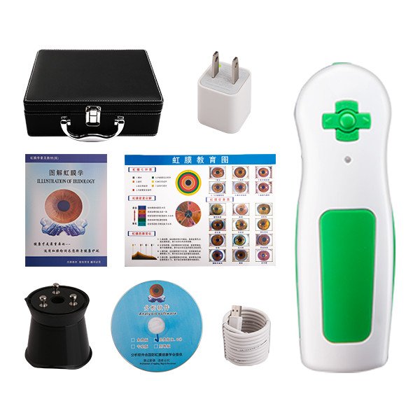
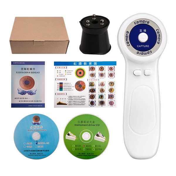
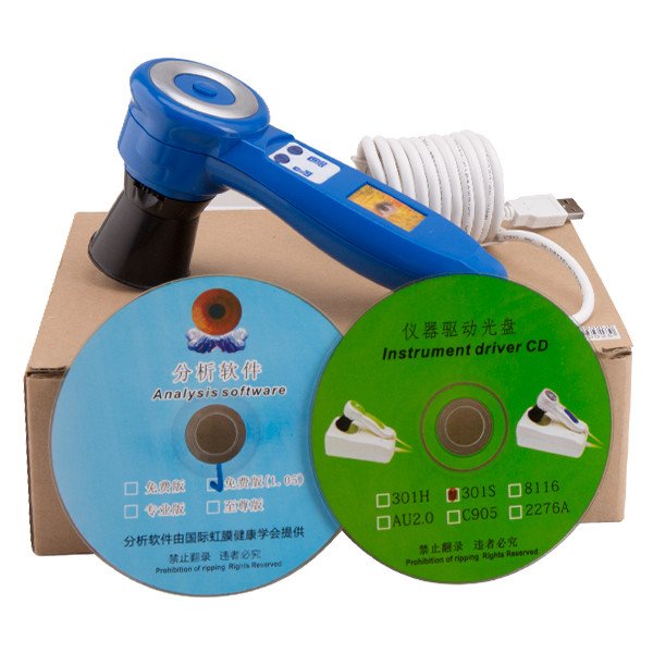
We supply two representative models you may see in supplier material and sample requests: MAIKONG MK 980U and MK-9822U. Below we list the verified model facts you should rely on when comparing example images and capture claims.
| Model | Sensor / Max Resolution | Illumination | Software / Languages | Supported OS | MOQ / Lead time / Warranty | Declared certification |
|---|
Common mistakes and image-quality issues to watch for when importing or reselling iris equipment
When we evaluate supplier examples we look for recurring problems importers should flag:
- Over-processed marketing images: heavy sharpening, contrast or non-representative background that hide optical limitations.
- Missing metadata: inability to link an exported analysis to the raw capture erodes trust in software claims.
- Uneven illumination and reflections: visible LED hot spots, specular reflections or shadowing across the iris indicate capture set-up problems or a misaligned illuminator.
- Insufficient magnification: a “high-res” image that lacks detail in magnified sectors suggests interpolation rather than true sensor resolution.
How comparable provider software affects example outputs and multilingual support considerations
Software governs both how images are stored and what annotated exports look like. For buyers in Canada a few practical checks reduce integration friction:
- Ask for images exported from the vendor software you will receive. Confirm the software version and included languages. (For example, MAIKONG lists MaikongCVAdvance English/Spanish/French/German/Hungarian/Italian/Japanese/Persian for MK-9822U; preserve that exact listing when you request samples.)
- Confirm file export formats (TIFF, JPEG, or proprietary database) and whether a software license is tied to a USB key or user account.
- Request a sample analysis report exported to PDF so you can verify fonts and language support for client-facing materials in Canada (English/French as needed).
Limitations: what comparable provider images do not demonstrate (clarifying medical boundaries)
comparable provider images only show external ocular features and software overlays. They do not provide medical diagnoses, laboratory correlation, or clinical outcomes. We will not and do not claim that images prove health conditions. Any clinical use must comply with local regulations and be carried out by appropriately trained practitioners.
Checklist for importers: how to request example images and evaluation files from suppliers
Use this checklist when you request samples — it shortens evaluation time and avoids back-and-forth.
- State models: e.g., MK 980U (5MP CCD) or MK-9822U (12 MP) and include data-fact-id where available.
- Request file types: 1–3 TIFFs (left/right/magnified), 1 JPEG for listing, and one PDF export from analysis software.
- Ask for capture metadata: timestamp, LED mode, lens used, software version.
- Require at least one unedited sample that includes the raw EXIF and one annotated export for training materials.
- Specify the intended use (clinic records, training, online listing) and required languages for software or exported reports.
Suggested internal workflow: accept, test, document and approve supplier image examples before listing
We recommend a short, repeatable evaluation protocol you can run in-house or through a local partner:
- Open raw files on a neutral workstation and confirm resolution, EXIF data and that the image matches the supplied annotation.
- Compare paired illumination modes to confirm LED control and note any reflections or hot spots.
- Measure effective detail: crop a magnified sector and inspect for pixelation at intended display/output sizes.
- Verify software export: open the supplier PDF report and confirm language support and accurate mapping to the raw image.
- Approve or reject for listing; if rejected, return clear, itemised feedback (e.g., “provide raw TIFF with EXIF and magnified sector shot with iris LED mode”).
Useful internal links and visual reference
For training and reference materials see our pages on comparable provider Course, general comparable provider guidance, and the comparable provider chart how to read resource.
One practical next step (how to request samples from us)
If you are ready to evaluate image examples for a Canadian distribution or OEM opportunity, email our sourcing contact with the following details: intended use (clinic / training / retail listing), preferred model(s) (e.g., MK 980U or MK-9822U), and the sample types you require (left/right iris TIFF, magnified sector, sclera and software-exported PDF). Send requests to Lucy@myiridology.us or WhatsApp +86 135-1090-7401.
Request a tailored sample pack
Please include the model names, the sample file list you need, and whether bilingual (English/French) exports are required. Pricing and configuration quotes are available on inquiry.
Important Disclaimer
Content is for educational and informational purposes only and does not constitute medical advice, diagnosis, or treatment. comparable provider is a complementary wellness practice and should not replace professional medical evaluation. Our comparable provider cameras and iriscopes are professional wellness imaging tools; users should receive appropriate training and comply with applicable local laws and professional requirements in Canada.
This content is provided for learning, information sharing and professional communication only. It is not medical advice and is not a basis for treatment or diagnosis. Confirm suitability, operation and purchasing requirements against the product manual, local regulations and qualified professional guidance.
Contact us online by Whatsapp:



Maybe you like also
Copy©2026-2027