
Iridology Case Studies of Lesions in Iris Closing: Understanding the Evidence, Boundaries, and Professional Practice Standards
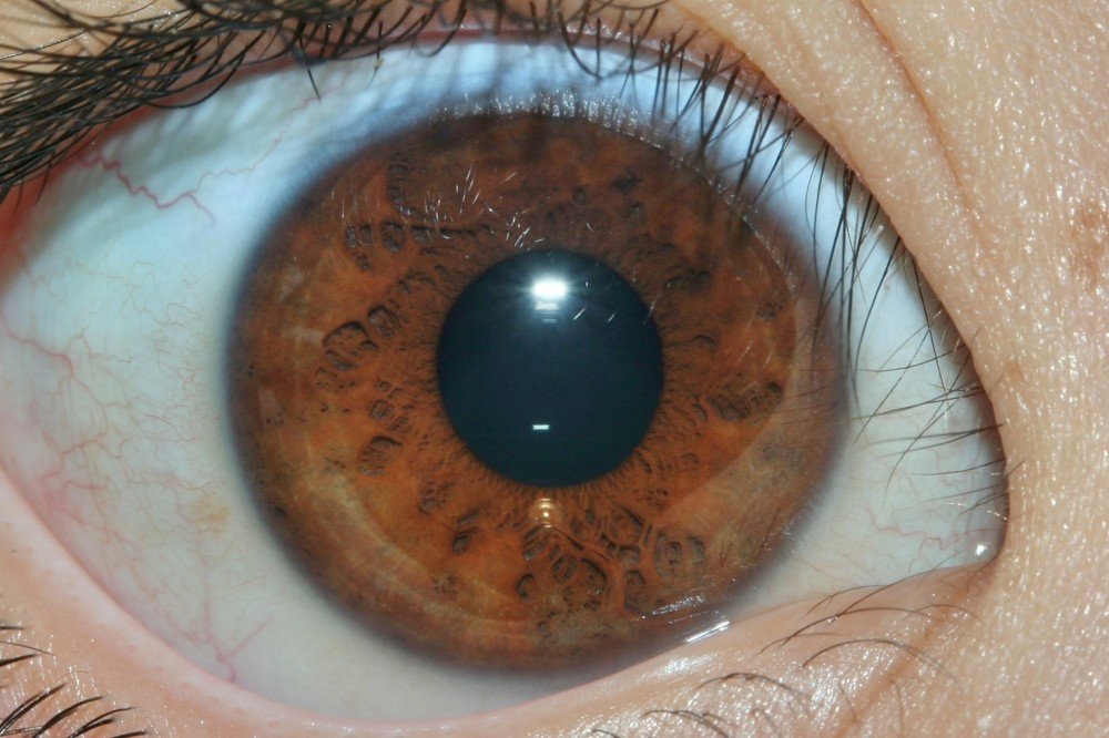
When health professionals search for iridology case studies of lesions in iris closing, they’re asking a critical question: what does documented evidence actually show about iris observation in traditional health practice? As someone who has worked alongside iridology practitioners, equipment manufacturers, and natural health clinics for years, I understand this question reflects both professional curiosity and practical concern about investing in equipment, training, or practice integration.
The phrase “lesions in iris closing” sits at the intersection of traditional iridology terminology and medical ophthalmology language, creating confusion that deserves careful clarification. In medical science, an iris lesion refers to structural abnormalities like iris melanoma, iris nevus, or other pathological changes requiring immediate ophthalmology referral. In traditional iridology practice, practitioners use “lesion” to describe constitutional markings, fiber patterns, or pigmentation changes they believe correlate with body system function over time.
This article provides honest, evidence-based examination of what case documentation in iridology actually shows, where scientific validation exists and where it doesn’t, and how professionals can make informed decisions about equipment, practice boundaries, and client communication. I’ll address the core questions: Is there iridology evidence supporting these observations? What does iridology research actually demonstrate? And critically, where are the boundaries that separate traditional observation from medical diagnosis?
Medical Disclaimer: This content discusses traditional iridology observation for educational and complementary health reference only. Iridology case studies described here document practitioner observations within traditional iridology frameworks and do not constitute medical diagnosis, treatment, or disease detection. Any iris changes, lesions, or abnormalities require evaluation by licensed ophthalmologists or medical doctors. This information does not replace professional medical examination.
What Does “Lesion Closing” Mean in Traditional Iridology vs. Medical Ophthalmology?
The terminology confusion between iridology case studies of lesions in iris closing and actual medical pathology creates the first major challenge for practitioners trying to establish credible practice. Let me clarify these completely different frameworks.
Medical Ophthalmology Definition of Iris Lesions
In evidence-based ophthalmology, iris lesions are structural abnormalities visible during dilated fundus examination and anterior segment examination. According to data from ocular oncology services, the most commonly documented iris lesions include:
- Iris melanoma – malignant tumor requiring immediate intervention, accounting for approximately 3-10% of uveal melanomas
- Iris nevus – benign pigmented lesion requiring monitoring for transformation signs over years
- Iris cysts – fluid-filled structures in anterior chamber or posterior iris surface
- Inflammatory lesions – associated with uveitis, secondary glaucoma, or ectropion uveae
- Vascular abnormalities – neovascularization of iris tissue indicating systemic or ocular disease
These medical conditions require slit lamp examination, sometimes ultrasound biomicroscopy, and regular ophthalmology follow-up. The concept of a lesion “closing” in medical terms would refer to resolution of inflammation, surgical repair, or in tragic cases, progression to more serious pathology. Medical documentation uses standardized protocols, measurements in millimeters, photographic comparison over months or years, and correlation with systemic health markers.
Traditional Iridology Definition of “Lesions” and “Closing”
In traditional iridology practice—developed primarily in 19th and 20th centuries by practitioners like Bernard Jensen and others—the term “lesion” refers to something entirely different. Iridology practitioners observe and document:
- Constitutional fiber patterns – the radial arrangement and density of iris fibers
- Pigmentation variations – color deposits, spots, or rings in specific iris zones
- Lacunae and crypts – natural fiber separations or spaces in iris structure
- Contraction furrows – circular grooves in iris tissue
- Nerve rings – concentric circular patterns
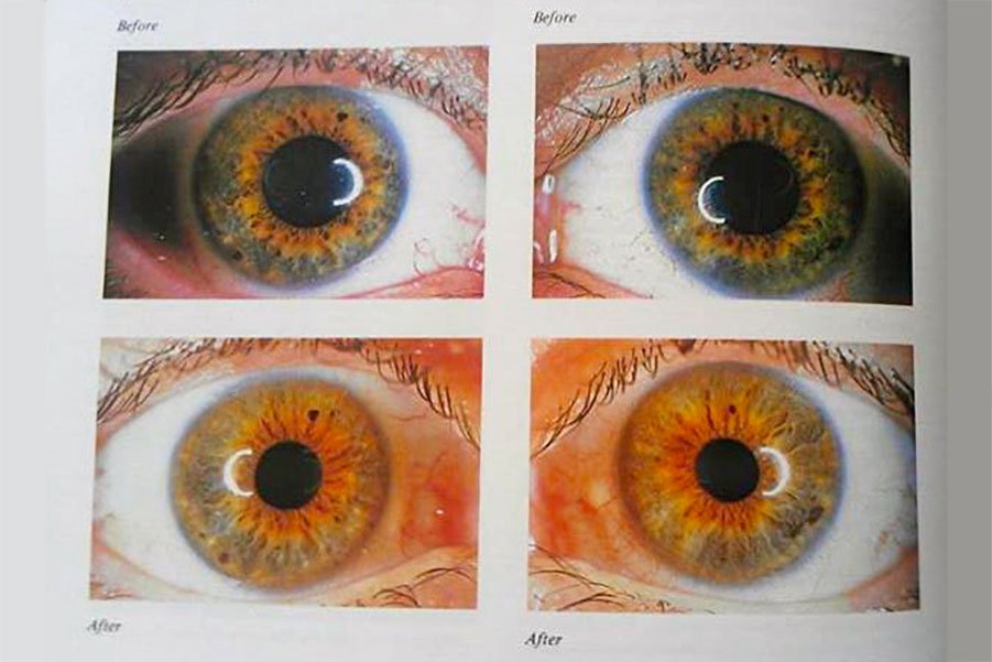
When iridology practitioners speak of a “lesion closing,” they typically mean observational changes in these constitutional markings over time—perhaps fiber patterns appearing denser, pigmentation spots fading, or lacunae appearing less pronounced. These observations are recorded through iridology camera photography at intervals ranging from weeks to years.
Critical Boundary: Traditional iridology “lesions” and medical iris lesions are not the same thing. Iridology observations of fiber patterns or pigmentation should never be confused with, or used to assess, actual pathological iris lesions like melanomas, nevi requiring monitoring, or inflammatory conditions. Any suspicious iris change must be referred immediately to ophthalmology.
The Documentation Challenge in Iridology Case Studies
When examining iridology case studies with pictures, the challenge becomes separating normal aging changes in iris appearance, photographic lighting variations, and camera resolution differences from genuine pattern changes that practitioners interpret through their traditional frameworks.
Legitimate case documentation requires:
Technical Standards
- Consistent lighting conditions across all images
- Minimum 12-megapixel resolution for fiber detail
- Standardized distance and focus protocols
- Same equipment throughout case timeline
- Precise timestamp and patient data management
Observational Standards
- Documented practitioner observations at each session
- Client-reported changes in subjective wellbeing
- No claims of diagnosing specific diseases
- Clear notation when medical referral made
- Acknowledgment of observation limitations
Without these standards, iridology case studies become anecdotal photography collections rather than systematic observation records. For professionals considering iridology camera for sale options, understanding these documentation requirements shapes equipment selection decisions.
The Historical Development of Iridology Case Documentation
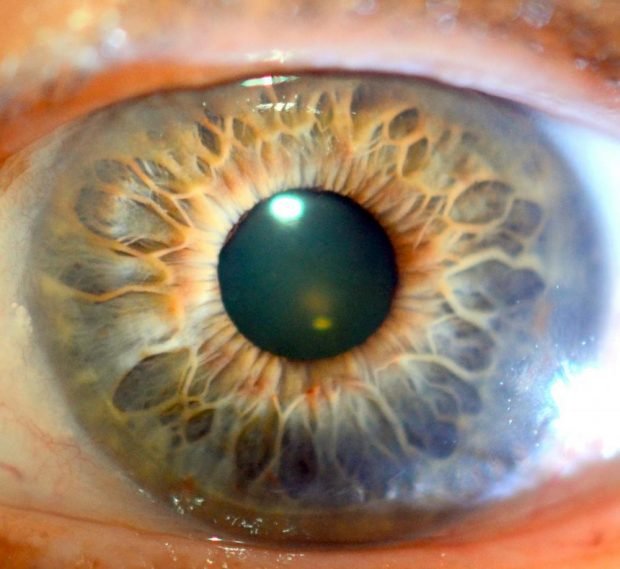
Understanding the evolution of iridology case studies of lesions in iris closing requires examining how this practice developed over approximately 150 years, from subjective observation to modern digital documentation attempts.
19th Century Origins: Ignatz von Peczely and Early Observations
The commonly cited origin story involves Hungarian physician Ignatz von Peczely, who in the 1860s reportedly observed iris changes in an owl with a broken leg. Whether historically accurate or apocryphal, this narrative established iridology’s foundational premise: that iris markings might correlate with body conditions elsewhere.
Early practitioners had no photographic equipment for case documentation. “Case studies” consisted of:
- Hand-drawn iris diagrams with practitioner annotations
- Written descriptions of fiber patterns and colorations
- Subjective practitioner memory across patient visits
- No standardized measurement or comparison protocols
This era established iridology terminology still used today but provided no rigorous evidence base that would satisfy modern scientific standards.
Early-to-Mid 20th Century: Bernard Jensen and Systematic Charting
American chiropractor Bernard Jensen became iridology’s most influential modern proponent, developing the widely-used Bernard Jensen iridology chart that maps body organs and systems to specific iris zones. Jensen documented thousands of patient observations across his decades of practice.
However, Jensen’s case documentation methods had significant limitations:
What Jensen’s Work Provided
- Systematic framework for consistent observation
- Standardized iris zone mapping used globally
- Extensive written case descriptions
- Integration with nutritional health approaches
- Training methodology for teaching practitioners
What Jensen’s Work Lacked
- Controlled studies with comparison groups
- Blinded assessment protocols
- Statistical analysis of observation accuracy
- High-resolution photographic documentation
- Peer review by medical research community
Jensen’s work established iridology as a structured practice system but did not provide the kind of evidence that would establish iridology proof within medical science frameworks.
Late 20th Century: Attempts at Scientific Validation
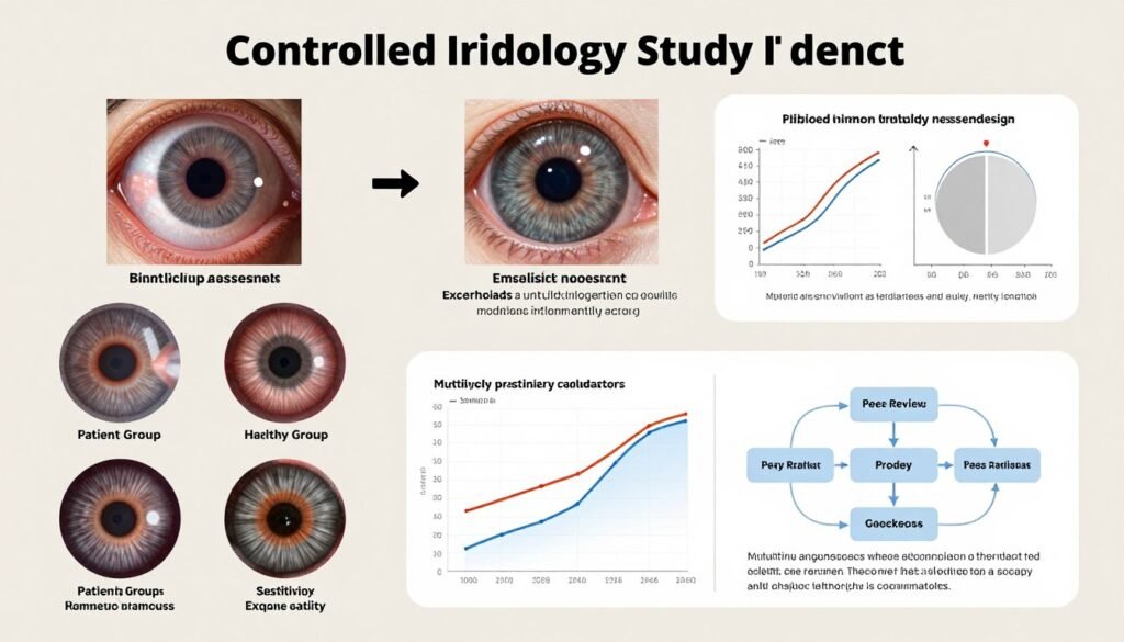
As evidence-based medicine became the standard, researchers attempted to test iridology claims systematically. Several key studies examined whether iridologists could accurately identify health conditions from iris photographs:
| Study Year | Research Design | Sample Size | Key Findings | Conclusion |
| 1979 | Blinded iris photo assessment | 143 patients | Iridologists performed no better than chance at identifying kidney disease from iris photographs | No validation of diagnostic claims |
| 1988 | Controlled evaluation of gallbladder disease detection | 39 patients | No correlation found between iris markings and confirmed gallbladder pathology | Failed to support diagnostic capability |
| 2000 | Systematic review of iridology research literature | Meta-analysis | Concluded available evidence does not support use of iridology as diagnostic tool | Insufficient scientific support |
| 2005 | Kidney dysfunction detection study | 110 participants | Iridologists showed poor sensitivity and specificity for detecting kidney problems | Not useful for screening purposes |
These studies, while limited in scope, consistently failed to validate iridology accuracy claims for specific disease diagnosis. This research forms the basis for critics’ assertions about iridology quackery. However, iridology proponents argue these studies tested diagnostic claims rather than the broader constitutional assessment and wellness monitoring framework many modern practitioners actually use.
21st Century: Digital Photography and Software Documentation
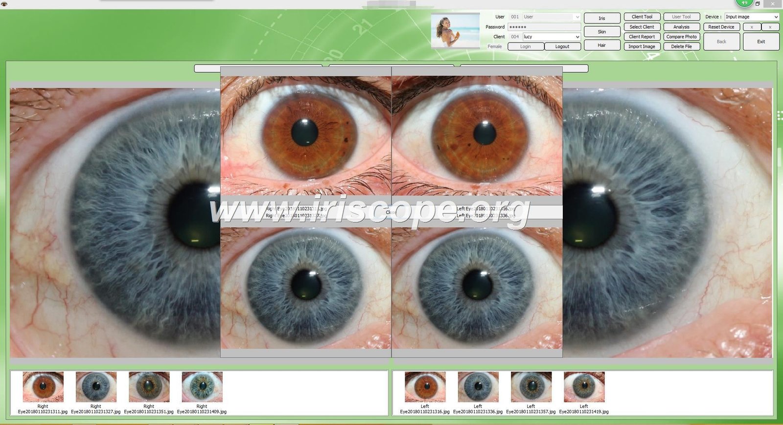
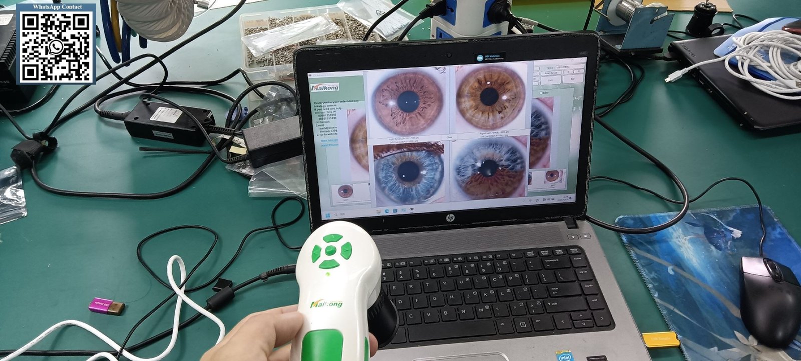
Modern digital technology transformed case documentation possibilities. Today’s practitioners use high-resolution iriscope systems with specialized iridology software enabling:
- 12+ megapixel iris photography capturing fine fiber detail
- Consistent LED lighting eliminating illumination variables
- Software comparison tools for side-by-side image analysis across months or years
- Digital archiving with precise timestamps and client data management
- Image enhancement features revealing subtle pattern changes
This technological capability means contemporary iridology case studies of lesions in iris closing can achieve photographic documentation quality impossible in earlier eras. However, improved image quality does not resolve the fundamental question of whether observed iris changes have the health correlations practitioners claim.
Documented Case Studies: What Can Actually Be Observed and Recorded?
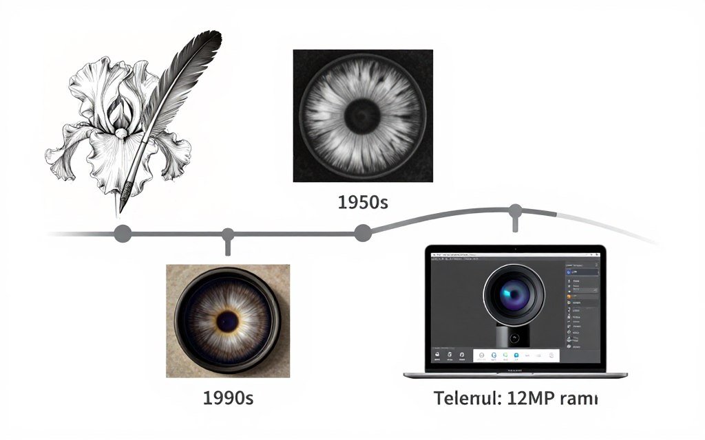
When examining legitimate iridology case studies with pictures, we must distinguish between what can be objectively photographed and what practitioners interpret through their traditional frameworks.
Observable and Documentable Iris Changes Over Time
Certain iris characteristics genuinely do change across months or years, and these changes can be photographed consistently with proper equipment. Objectively documentable observations include:
Pigmentation Changes
Iris color can subtly shift with age, medication use, or certain systemic conditions. Pigment deposits may appear, darken, or occasionally lighten. These changes are photographically verifiable with consistent lighting and equipment.
Pupil Response Variations
While not typically part of static iridology photography, pupil size and reactivity can change with neurological function, medication, or systemic health. Dynamic pupillometry requires specialized equipment beyond standard iridology cameras.
Vascular Pattern Changes
Blood vessel patterns in iris tissue can become more or less visible over time, particularly with cardiovascular changes or inflammatory conditions. High-resolution photography documents these vascular changes.
However, documenting that these changes occur does not automatically validate iridology interpretations about what these changes mean regarding body system function or health status. This represents the core challenge in evaluating iridology evidence.
Typical Iridology Case Study Structure
Professional practitioners using systematic documentation typically structure case observations as follows:
Initial Session Documentation: Baseline iris photography of both eyes, practitioner observations of fiber patterns, constitutional type assessment per traditional iridology charts, client health history and current concerns, lifestyle and nutritional factors
Follow-up Session Documentation: New iris photography using identical equipment and lighting, side-by-side comparison with previous images, practitioner observations of any pattern changes, client-reported changes in subjective wellbeing, documentation of any lifestyle or health interventions between sessions
This protocol allows practitioners to create iridology case studies showing temporal progression. What remains unproven is whether practitioner interpretations of observed changes have valid correlation with actual physiological changes in body systems.
Example Case Documentation: Constitutional Markings Over Time
A typical documented observation might proceed as follows:
Case Background: Female client, age 42, reports persistent digestive discomfort. Initial iris examination shows pronounced lacunae (fiber separations) in the intestinal zone according to traditional iridology chart mapping (approximately 6-7 o’clock position in right iris). Practitioner documents these observations alongside client’s symptoms.
Intervention Period: Client works with nutritionist for dietary modifications, addresses food sensitivities, increases fiber and probiotic intake over six months. Regular medical gastroenterology workup rules out serious pathology.
Follow-up Documentation: Six months later, new iris photography appears to show the previously noted lacunae appearing less pronounced, with surrounding fiber patterns appearing slightly denser. Client reports subjective improvement in digestive comfort.
Practitioner Interpretation: Traditional iridology framework suggests fiber pattern changes correlate with improved digestive function. Practitioner documents this as “lesion closing” or constitutional strengthening in that iris zone.
Scientific Limitation: This documentation cannot establish causation. The client made multiple interventions, experienced natural symptom fluctuation over time, and iris photography variables (lighting, focus, client’s pupil size that day) may contribute to apparent visual differences. The correlation between iris pattern changes and digestive improvement remains unproven.
This example illustrates why iridology research paper attempts struggle to establish causation even when correlation seems present. Multiple confounding variables prevent clean scientific conclusions.
Limitations in Current Case Study Documentation
Even the most professionally documented iridology case studies of lesions in iris closing face inherent methodological limitations:
| Limitation Type | Description | Impact on Evidence Quality |
| Observer Bias | Practitioners expecting to see improvement may unconsciously perceive patterns as changed when differences are minimal or photographic artifacts | Reduces objectivity of assessments |
| Selection Bias | Published case studies typically showcase apparent successes rather than cases showing no iris pattern changes despite health improvements | Creates false impression of consistency |
| Photographic Variables | Even with good equipment, subtle lighting, focus, or pupil dilation differences between sessions can create apparent pattern changes that aren’t physiological | Cannot distinguish real from artifactual changes |
| Lack of Control Groups | Case studies don’t compare iris changes in people receiving interventions versus those who don’t, making causation impossible to establish | Cannot prove intervention caused observed changes |
| Subjective Interpretation | Different practitioners viewing identical iris photographs may identify different “lesions” or interpret patterns differently | Poor inter-rater reliability |
| Temporal Ambiguity | Iris patterns do change naturally with aging; distinguishing age-related changes from health-related changes is problematic | Cannot isolate health correlations |
These limitations explain why organizations evaluating is iridology a science consistently conclude that current evidence does not support diagnostic or specific health assessment claims, even when practitioners can document interesting iris pattern observations over time.
Considering Professional Iridology Documentation Equipment?
If you’re establishing an iridology practice or integrating iris observation into your natural health clinic, equipment quality directly impacts documentation credibility. Our team provides consultation on evidence-based equipment selection that meets professional standards while acknowledging appropriate practice boundaries.
Scientific Research on Iridology: Current Evidence and Limitations
The question “is iridology legit” depends entirely on what claims are being evaluated. Let me break down what research on iridology actually shows versus what remains unproven.
What Scientific Research Has Attempted to Test
Most formal iridology research paper publications have focused on specific diagnostic claims—can practitioners identify particular diseases or organ dysfunction from iris photographs? These studies employ standard scientific methodology:
- Blinded assessment: Iridologists evaluate iris photos without knowing patients’ actual health status
- Control groups: Comparing patients with confirmed conditions against healthy controls
- Statistical analysis: Calculating sensitivity, specificity, and accuracy of practitioner assessments
- Multiple evaluators: Testing whether different practitioners reach similar conclusions from same photos
The consistent finding across these studies: iridologists performing diagnostic assessments showed accuracy no better than random chance. A comprehensive 2011 meta-analysis examining multiple controlled trials concluded there was insufficient evidence to support iridology as a diagnostic modality.
Why These Studies May Not Reflect Actual Practice
However, many contemporary practitioners argue these studies test claims they don’t actually make. Modern iridology science and practice increasingly focuses on:
What Research Tested
- Diagnosing specific diseases from single iris photo
- Identifying organ pathology without patient history
- Using iris markings as sole diagnostic tool
- Replacing conventional medical examination
What Many Practitioners Actually Claim
- Observing constitutional tendencies over time
- Integrating iris observation with comprehensive health assessment
- Using observations as complementary information
- Supporting wellness monitoring, not disease diagnosis
This gap between what research tested and what practitioners claim creates ongoing debate about iridology is it accurate. The studies convincingly refute diagnostic accuracy claims but don’t necessarily address whether iris observation provides value in broader wellness contexts.
Areas Where Some Research Shows Interesting Correlations
While diagnostic claims remain unvalidated, some research has identified intriguing correlations between iris characteristics and certain conditions:
| Research Area | Findings | Interpretation | Limitations |
| Iris Crypts and Connective Tissue | Some studies found correlation between number of iris crypts and joint hypermobility | May reflect shared genetic factors affecting connective tissue throughout body | Correlation doesn’t equal causation; clinical utility unclear |
| Arcus Senilis (Corneal Ring) | Well-established correlation with cholesterol levels and cardiovascular risk in younger patients | This is recognized in conventional medicine, not unique to iridology | Not traditionally part of iridology mapping system |
| Iris Color and Pain Sensitivity | Research suggests people with lighter iris colors may have slightly different pain thresholds | Genetic factors affecting melanin may influence neurological factors | Effect sizes small; not useful for clinical pain assessment |
| Pupillary Response and Autonomic Function | Pupil reactions can indicate autonomic nervous system function | Recognized in neurology; dynamic pupillometry has medical applications | Requires specialized equipment beyond static iris photography |
These findings suggest some iris characteristics do correlate with systemic factors, but they don’t validate the specific organ-mapping claims in traditional iridology chart how to read systems.
The “Iridology Quackery” Critique and Reasonable Responses
Medical skeptics using terms like iridology quackery point to:
Legitimate Scientific Concerns
- No anatomical pathway connecting specific body organs to specific iris zones
- Failed controlled studies of diagnostic accuracy across multiple decades
- Risk of delayed medical diagnosis if patients rely on iridology instead of appropriate testing
- Inconsistent practitioner interpretations of same iris photographs
- Lack of peer-reviewed research supporting specific health claims
Practitioner Counterarguments
- Constitutional observation differs from organ-specific diagnosis
- Practice has evolved beyond claims tested in older studies
- Used as complementary tool, not replacement for medical care
- Practitioner variation reflects interpretive art component
- Limited research funding prevents comprehensive study
Honest assessment requires acknowledging both perspectives. The scientific evidence does not support iridology as a diagnostic tool for specific diseases or organ pathologies. However, this doesn’t automatically invalidate all observation-based wellness practices when positioned appropriately within complementary health contexts rather than as medical diagnosis.
Understanding “Is Iridology Pseudoscience?”
The question “is iridology pseudoscience” depends on how specific practitioners present and use it:
Pseudoscientific positioning includes: Claiming to diagnose specific diseases without medical testing, asserting that iris markings definitively indicate organ dysfunction, promoting iridology as replacement for medical examination, making unsubstantiated health promises, ignoring contradictory research evidence.
Reasonable positioning includes: Presenting as traditional observation system for constitutional assessment, acknowledging lack of diagnostic validation, using as one tool among many in comprehensive health evaluation, emphasizing it cannot replace medical diagnosis, referring clients for appropriate medical testing, documenting observations without making disease claims.
Professionals asking “is iridology valid” must understand this distinction. The practice can have validity within properly bounded traditional observation contexts while lacking validity as a medical diagnostic tool.
Why Iridology Cannot Replace Medical Diagnosis: Understanding the Boundaries
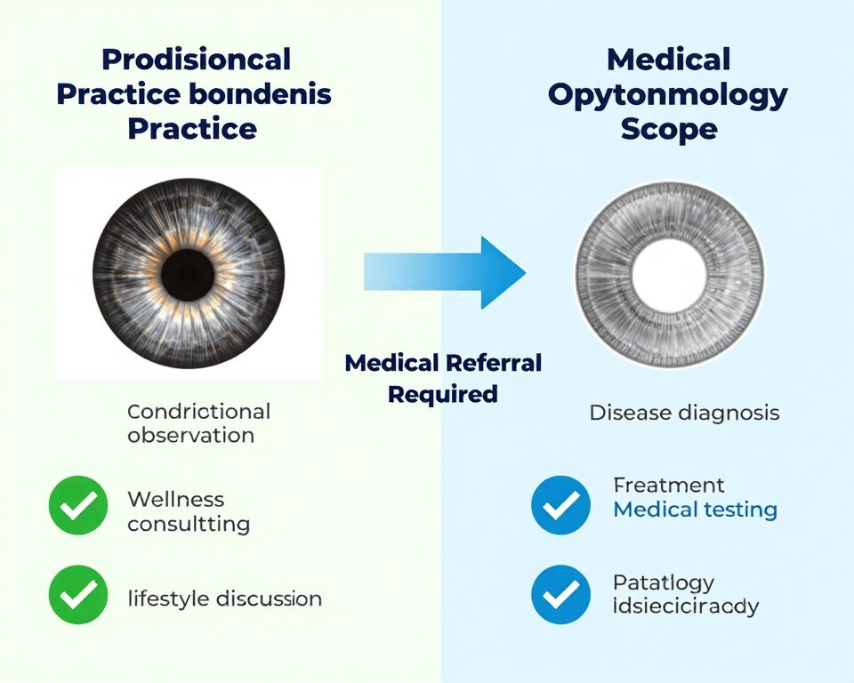
Perhaps the most important information for anyone researching iridology case studies of lesions in iris closing is understanding absolute boundaries that separate traditional observation from medical diagnosis and treatment.
Critical Medical Conditions Requiring Immediate Ophthalmology Referral
Any practitioner using iridology camera equipment must recognize when iris observations indicate potential serious pathology requiring immediate medical attention. These include:
Immediate Medical Referral Required For:
- Iris melanoma or suspicious pigmented lesions: Any new, growing, or irregular pigmented spot in iris tissue may represent malignancy requiring ocular oncology evaluation. Uveal melanoma represents serious cancer risk.
- Iris nevi showing growth or transformation: While many iris nevi are benign, those showing size increase, irregular borders, or associated symptoms require ophthalmology monitoring.
- Neovascularization (abnormal blood vessel growth): New blood vessels on iris surface often indicate serious underlying conditions like diabetic retinopathy, retinal vein occlusion, or ocular ischemia.
- Inflammatory signs: Iris tissue inflammation, hypopyon (layered white cells in anterior chamber), or associated eye pain may indicate uveitis, infection, or autoimmune conditions requiring urgent treatment.
- Structural abnormalities: Iris damage, dislocation, ectropion uveae (posterior iris pigment visible on anterior surface), or irregular pupil shape may indicate trauma, glaucoma, or other serious conditions.
- Secondary glaucoma indicators: Abnormal anterior chamber angle appearance or iris changes associated with elevated eye pressure require immediate evaluation.
Traditional iridology practitioners are not trained to differentiate these serious medical conditions from constitutional markings. This is why professional boundaries are non-negotiable—iris observation must remain separate from medical diagnosis.
What Medical Ophthalmology Provides That Iridology Cannot
Understanding iridology in medical science requires recognizing what actual ophthalmology examination includes:
| Medical Examination Component | What It Assesses | Why Iridology Cannot Replace It |
| Dilated Fundus Examination | Retina, optic nerve, blood vessels, macula for diseases like diabetic retinopathy, macular degeneration, retinal detachment, glaucoma damage | Iridology only photographs anterior iris surface; cannot see posterior eye structures where most serious diseases manifest |
| Intraocular Pressure Measurement | Eye pressure to detect glaucoma risk | Requires specialized tonometry equipment; iris appearance cannot indicate pressure levels |
| Slit Lamp Examination | Detailed biomicroscopy of cornea, iris, lens, anterior chamber structures | Medical-grade biomicroscope with multiple magnifications and specialized lighting techniques provides detail beyond consumer iridology cameras |
| Visual Field Testing | Peripheral vision loss indicating neurological or glaucoma issues | Functional testing of vision pathways; iris structure cannot predict visual field defects |
| Optical Coherence Tomography (OCT) | Cross-sectional retinal imaging showing layer structure and disease | Advanced imaging technology assessing posterior eye; completely different from iris photography |
| Gonioscopy | Anterior chamber angle structure to assess glaucoma risk and type | Requires specialized lens and technique; angle visualization crucial for glaucoma management |
This comparison illustrates why asking “is iridology accurate” for medical diagnosis is the wrong question—the tools assess fundamentally different things.
Legal and Ethical Practice Boundaries in United States
For professionals considering integrating iridology into US-based practice, understanding legal boundaries is essential:
What Is Generally Permitted (with appropriate disclaimers)
- Offering traditional iridology observation as complementary wellness assessment
- Documenting constitutional patterns according to traditional iridology frameworks
- Providing educational information about iridology history and traditional use
- Photographing iris structures for comparative observation over time
- Discussing lifestyle and nutritional approaches within practitioner’s scope of practice
What Is Prohibited or Legally Risky
- Claiming to diagnose medical diseases or conditions from iris observation
- Suggesting iridology can replace medical testing or diagnosis
- Prescribing medical treatment based solely on iris observations
- Using terms like “diagnosis,” “disease,” or “cure” in iridology context
- Delaying client’s pursuit of appropriate medical care
- Practicing beyond one’s professional license and training scope
State regulations vary significantly. Some states have specific rules governing alternative health practices, while others apply general medical practice laws. Consulting with healthcare attorney familiar with your state’s regulations is essential before establishing iridology services.
Informed Consent and Client Communication
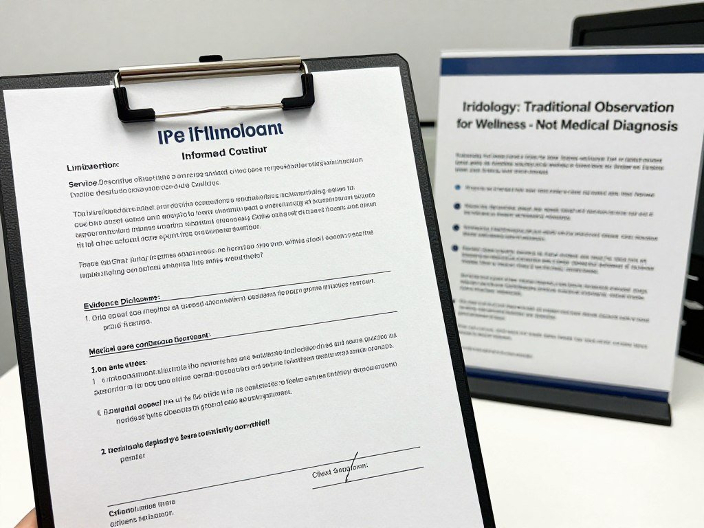
Professional practice requires clear communication about iridology boundaries. Effective informed consent should include:
“Iridology is a traditional observation system used in complementary health practice. It involves photographing and observing iris characteristics that practitioners correlate with constitutional tendencies and general wellness patterns. Iridology is not medical diagnosis and cannot detect diseases, replace medical examination, or substitute for appropriate medical testing. Any health concerns require evaluation by licensed medical professionals. The observations and suggestions provided are for educational and wellness purposes only.”
This transparency builds trust while protecting both practitioner and client. It also addresses the concerns behind searches like “iridology legit“—legitimacy comes from honest boundary-setting, not exaggerated claims.
When to Refer to Medical Professionals
Any iridology practitioner should immediately refer clients to ophthalmology or primary care when they observe or clients report:
Vision or Eye Symptoms
- Sudden vision changes or loss
- Eye pain, redness, or discharge
- Flashes of light or new floaters
- Halos around lights
- Double vision
- Any eye injury or trauma
Iris Appearance Concerns
- New or changing pigmented spots
- Visible blood vessels where none existed before
- Asymmetry between eyes
- Irregular pupil shape
- Apparent inflammation or tissue changes
- Any appearance suggesting pathology
Professional boundaries aren’t limitations—they’re the foundation of ethical, responsible practice that respects both traditional observation systems and evidence-based medicine.
Equipment Requirements for Credible Iridology Case Documentation
If you’re committed to systematic iridology case studies of lesions in iris closing documentation, equipment quality directly impacts observation credibility and professional practice standards. Let me outline what serious practitioners actually need.
Minimum Technical Specifications for Professional Documentation
Not all devices marketed as iridology camera for sale meet professional documentation standards. Minimum requirements include:
| Specification Category | Minimum Requirement | Professional Standard | Why It Matters |
| Image Resolution | 8 megapixels | 12+ megapixels | Captures fine fiber detail essential for documenting subtle pattern changes over time |
| Lighting System | Adjustable LED ring light | Adjustable LED with color temperature control | Consistent, shadow-free illumination eliminates lighting variables between sessions |
| Magnification | 20x-30x optical magnification | 30x+ with digital zoom capability | Reveals constitutional detail invisible to unaided eye; allows documentation of specific iris zones |
| Focus System | Manual focus with depth indication | Auto-focus with manual override and focus lock | Ensures consistent focal plane across all images in longitudinal case studies |
| Software Integration | Basic image capture and storage | Dedicated iridology software with comparison tools, client database, report generation | Enables systematic case management, side-by-side temporal comparison, and professional documentation |
| Connectivity | USB connection to computer | USB with multiple OS compatibility (Windows, Mac) | Ensures equipment works with practitioner’s existing computer systems and practice management software |
These specifications directly impact whether your iridology case studies will have sufficient image quality for credible documentation. Inadequate equipment produces images where apparent “pattern changes” may actually reflect resolution limitations or lighting inconsistencies rather than genuine iris changes.
Software Capabilities Supporting Professional Case Management
High-quality hardware requires equally capable iridology software for professional case documentation:
Essential Software Features
- Client database with comprehensive data fields
- Secure image storage with timestamp metadata
- Side-by-side comparison viewing across dates
- Annotation tools for marking observations
- Iridology chart overlay options
- Report generation with image integration
Advanced Professional Features
- Multi-language interface and reports
- Image enhancement and contrast adjustment
- Measurement tools for documenting lesion dimensions
- Historical trend tracking across multiple sessions
- HIPAA-compliant data security (for US practitioners)
- Integration with practice management systems
Future-Proof Capabilities
- Cloud backup and multi-device synchronization
- Software updates and feature additions
- Cross-platform compatibility (Windows and Mac)
- Technical support and training resources
- Customization for specific practice needs
- Integration of educational materials
When evaluating best iridology camera options, software capabilities often matter as much as hardware specifications. A 12MP camera with inadequate software provides less professional value than an integrated system with comprehensive case management tools.
Why Many Consumer-Grade Devices Are Insufficient
Practitioners sometimes ask whether smartphone macro lenses or basic USB cameras can serve for iridology case studies of lesions in iris closing. While these might capture iris images, they typically lack:
- Consistent lighting geometry: Built-in ring lights eliminate external lighting variables that change between sessions
- Standardized positioning: Purpose-built iriscopes maintain consistent distance and angle to iris
- Sufficient magnification: General-purpose cameras cannot achieve 30x magnification needed for constitutional detail
- Professional documentation software: Generic photo apps don’t provide systematic case comparison and management
- Practice credibility: Professional equipment signals commitment to systematic observation standards
The iriscope price difference between consumer and professional equipment reflects these capability differences. For practitioners building serious case study documentation, professional-grade systems represent necessary investment rather than optional upgrade.
MAIKONG Manufacturing Standards for Professional Equipment
At MAIKONG, we manufacture iriscope systems specifically designed for professional case documentation, understanding that equipment quality directly impacts practice credibility. Our MK-series devices incorporate specifications developed through 20 years of working with practitioners worldwide.
Here are the technical specifications of our current professional iridology camera systems:
| Model Series | Image Sensor | Resolution | Magnification Range | Lighting System | Software Compatibility |
| MK-9822U Standard | 12.0 MP CCD | 4032 x 3024 pixels | 30x optical | Adjustable LED ring (6-level) | Windows XP/7/8/10/11, Mac OS |
| MK-9822U Professional | 16.0 MP CMOS | 4608 x 3456 pixels | 30x optical + 2x digital | LED ring with color temperature adjustment | Windows XP/7/8/10/11, Mac OS |
| MK-9822U Advanced | 20.0 MP CMOS | 5472 x 3648 pixels | 40x optical + 4x digital | Dual-mode LED with polarization | Windows XP/7/8/10/11, Mac OS |
Each system includes comprehensive iridology software supporting 17 languages (English, Spanish, French, German, Italian, Portuguese, Russian, Polish, Czech, Bulgarian, Korean, Japanese, Persian, Simplified Chinese, Traditional Chinese, and Malaysian), enabling practitioners to serve diverse patient populations and produce reports in clients’ native languages.
Standard Package Inclusions for Professional Practice
Complete MAIKONG professional systems include all components needed for immediate case documentation implementation:
| Component | Specification | Quantity | Purpose |
| Iriscope Handset | 12/16/20 MP depending on model | 1 unit | Primary imaging device |
| 30x Iris Lens | Professional optical grade | 1 unit | High magnification iris visualization |
| Professional Carrying Case | Protective foam-lined case | 1 unit | Equipment protection and portability |
| USB Connection Cable | 1.5 meter shielded cable | 1 unit | Computer connectivity |
| Lens Protective Cover | Dust-proof cap | 1 unit | Lens protection during storage |
| Iridology Analysis Software | 2024 version, 17 languages | CD/Download | Case management and documentation |
| Encryption USB Dongle | Hardware security key | 1 unit | Software activation and licensing |
| Iridology Reference Chart | Laminated full-color chart | 1 set | Quick reference during consultations |
| User Manual | Comprehensive illustrated guide | PDF + Video tutorials | Setup, operation, and troubleshooting |
| Warranty Documentation | 1-year manufacturer warranty | Certificate included | Quality assurance and support |
This complete package approach ensures practitioners have everything needed for professional iridology case studies without requiring additional component purchases.
OEM and ODM Customization for US Distributors
For health equipment distributors, medical device importers, or established wellness brands seeking private-label iridology camera solutions, MAIKONG offers comprehensive customization services:
Hardware Customization (OEM)
- Model designation with your brand identifier
- Custom external housing colors and finish
- Branded laser engraving on device body
- Custom carrying case with your logo
- Modified packaging design and materials
- Specialized component configurations
Software Customization (ODM)
- Custom software interface with your branding
- Modified language selections for target markets
- Integration of your product recommendations
- Custom report templates with your logo
- Specialized features for your practice methodology
- Integration of educational content library
With manufacturing capacity supporting orders from single units to bulk wholesale quantities, and US warehouse stock enabling faster delivery to American markets, MAIKONG serves both individual practitioners and large-scale distributors. Our experience providing equipment to over 1,000 practitioners and companies globally—including major direct sales organizations, health supplement companies, and natural medicine clinics—demonstrates capability to support diverse business models.
Iridology Accuracy: What Studies Have Actually Measured
The search query “iridology is it accurate” reflects genuine concern about reliability before investing time, money, and professional reputation into practice integration. Let’s examine what accuracy actually means in this context and what research has measured.
Defining “Accuracy” in Different Contexts
The question of iridology accuracy becomes clearer when we distinguish what type of accuracy we’re discussing:
| Accuracy Type | Definition | Research Findings | Practical Implications |
| Diagnostic Accuracy | Ability to correctly identify presence or absence of specific diseases from iris observation alone | Multiple controlled studies show no better than chance accuracy; failed to meet clinical diagnostic standards | Iridology should not be positioned or used as diagnostic tool |
| Observational Consistency | Agreement between different practitioners observing same iris photographs (inter-rater reliability) | Poor to moderate consistency; different practitioners often identify different patterns or interpret same patterns differently | Subjective interpretation component limits standardization |
| Temporal Documentation Accuracy | Ability to consistently photograph same iris features across multiple sessions for comparison | With proper equipment and protocols, photographic documentation can be highly consistent | Quality equipment and standardized protocols enable reliable observation records |
| Pattern Recognition Accuracy | Ability to consistently identify specific iris features (lacunae, crypts, pigmentation, etc.) | Trained practitioners show moderate to good consistency in identifying structural features | Feature identification possible; health correlation interpretation remains unvalidated |
This breakdown shows that iridology can achieve accuracy in photographic documentation and basic feature identification while lacking accuracy in disease diagnosis or specific health condition prediction.
What Controlled Studies Actually Tested
When examining iridology research, understanding study design reveals what was actually measured:
Classic Diagnostic Accuracy Study Example
A typical controlled study design works as follows:
- Subject Selection: Researchers recruit patients with confirmed kidney disease (verified through medical testing) and healthy control subjects with normal kidney function
- Blinding Protocol: Iris photographs taken of all subjects without identifying information
- Practitioner Assessment: Experienced iridologists examine photos and indicate which subjects they believe have kidney disease based on iris markings in kidney zones per traditional iridology chart mapping
- Statistical Analysis: Researchers calculate sensitivity (percentage of actual disease cases correctly identified), specificity (percentage of healthy subjects correctly identified), and overall accuracy
Results consistently showed iridologist assessments performed at or near chance levels—essentially equivalent to random guessing. This does not mean practitioners saw nothing in the iris, but rather that what they observed didn’t reliably correlate with actual kidney disease presence.
Why These Studies May Not Reflect Modern Practice Claims
Many contemporary practitioners argue these studies don’t test what they actually claim. Modern iridology science and practice increasingly emphasizes:
What Studies Tested
- Single-time-point diagnosis from photograph only
- Specific disease presence/absence determination
- Organ-specific pathology identification
- Assessment without client history or other information
- Replacement of medical diagnostic testing
What Modern Practitioners Claim to Offer
- Constitutional tendency assessment over time
- General wellness pattern observation
- Holistic health evaluation integration
- Assessment within comprehensive consultation context
- Complementary information alongside medical care
This distinction matters when evaluating whether is iridology valid within properly bounded contexts. Research definitively shows it’s not valid for disease diagnosis, but hasn’t comprehensively tested broader wellness observation claims.
Factors Affecting Practical Accuracy in Case Documentation
For practitioners conducting longitudinal iridology case studies of lesions in iris closing, several factors affect documentation reliability:
This analysis reveals that technical documentation aspects (equipment, lighting, protocols) can achieve high reliability, while interpretive and correlation aspects remain problematic from scientific validity perspective.
Improving Documentation Reliability in Your Practice
Practitioners committed to highest possible accuracy in iridology case studies should implement these evidence-based improvements:
- Equipment standardization: Use identical USB iridology camera equipment for all sessions with same client
- Lighting protocols: Document specific lighting settings and maintain consistency across all photography sessions
- Positioning consistency: Establish and document standardized distance, angle, and focus settings
- Time-of-day consideration: Schedule follow-up sessions at similar times of day to minimize circadian pupil size variations
- Medication documentation: Note any medications affecting pupil dilation or iris appearance
- Blind assessment trials: Periodically have practitioners evaluate images without knowing session sequence to test observation consistency
- Client correlation tracking: Document client-reported symptoms and changes alongside iris observations without claiming causation
- Limitation acknowledgment: Explicitly note factors that could affect apparent iris changes (lighting, resolution, focus, aging, etc.)
These practices maximize the technical accuracy achievable while honestly acknowledging interpretive limitations—the hallmark of professional, ethically bounded iridology practice.
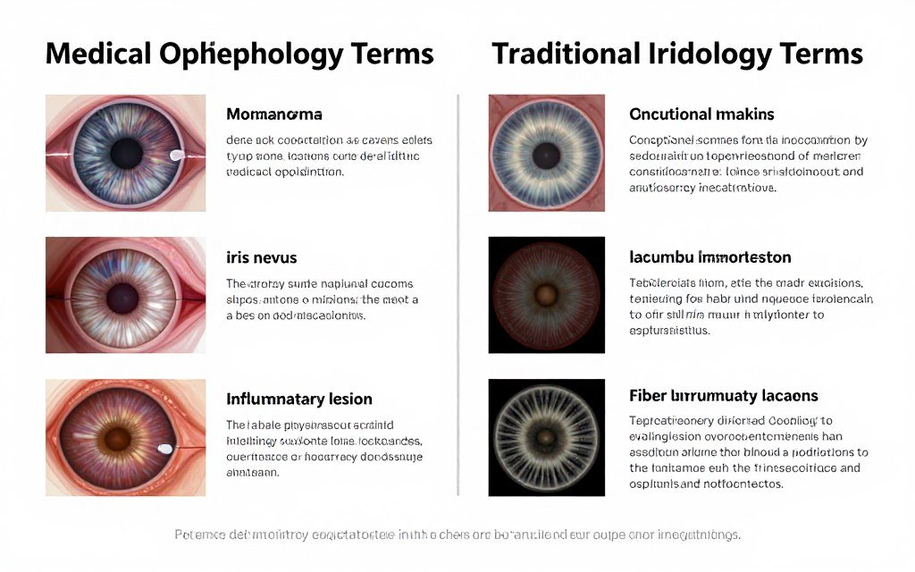
Common Misunderstandings: Iridology Terminology vs. Medical Pathology
One of the most dangerous areas of confusion surrounding iridology case studies of lesions in iris closing involves terminology overlap between traditional iridology language and actual medical pathology terms. This confusion can lead to delayed medical care or inappropriate practice claims.
Terms That Mean Different Things in Each Context
Several terms used in both iridology and ophthalmology have completely different meanings, creating dangerous potential for misunderstanding:
| Term | Iridology Meaning | Medical Ophthalmology Meaning | Why Confusion Is Dangerous |
| Lesion | Constitutional marking, fiber pattern variation, or pigmentation pattern interpreted as reflecting body system tendencies | Actual tissue abnormality, growth, or pathological change requiring medical evaluation (e.g., iris melanoma, iris nevus, inflammatory lesion) | Clients may believe iridology “lesion” assessment addresses medical concern about actual iris pathology, delaying ophthalmology referral |
| Inflammation | Traditional interpretation of certain fiber patterns or color variations as suggesting inflammatory tendencies in mapped body systems | Actual inflammatory process in eye tissue (uveitis, iritis) causing pain, photophobia, and requiring anti-inflammatory treatment | Actual ocular inflammation requires urgent medical treatment; confusing iridology interpretation with medical inflammation could delay care for serious condition |
| Healing Sign | Practitioners’ interpretation that iris patterns show “healing” or improvement in constitutional markings over time | Observable resolution of actual pathology (e.g., clearing of inflammatory cells, reduction in vascularization, lesion regression) documented through medical examination | Clients may believe iridology “healing signs” indicate actual disease resolution, potentially discontinuing necessary medical treatment |
| Acute vs. Chronic | Different fiber patterns or marking characteristics interpreted as reflecting acute versus chronic constitutional tendencies | Actual disease timeline—acute refers to sudden onset conditions requiring immediate intervention; chronic refers to long-standing conditions | Acute medical conditions require immediate attention; confusing iridology interpretation of “chronic” patterns with actual disease status could delay emergency care |
| Toxic Accumulation | Practitioners’ interpretation of certain pigmentation or pattern characteristics as reflecting “toxin” accumulation in body systems | No equivalent concept; actual toxic exposures affecting eyes cause specific medical conditions (e.g., drug toxicity, chemical burns) requiring treatment | Focuses attention on unproven “detoxification” instead of addressing actual health issues through evidence-based care |
| Degeneration | Interpretation of iris fiber pattern looseness or separation as suggesting degenerative tendencies | Actual tissue degeneration like macular degeneration, requiring ophthalmology care and affecting vision | Actual degenerative eye diseases require medical monitoring and treatment; iridology observations cannot diagnose or monitor these conditions |
This terminology confusion is why professional iridology practice requires extremely careful language choices and consistent reminders that observations relate to traditional constitutional frameworks rather than medical diagnosis.
What “Iris Closing” Actually Means in Each Context
The specific phrase “lesions in iris closing” from our focus keyword deserves particular attention:
Iridology Interpretation of “Closing”
In traditional iridology case studies, practitioners use “closing” to describe their observation that constitutional markings—particularly lacunae (fiber separations) or crypts—appear less pronounced or seem to “fill in” over time. They interpret this as positive change indicating:
- Improved constitutional strength in affected body system zones
- Response to nutritional or lifestyle interventions
- Enhanced vitality and wellness patterns
- Positive health trajectory indicators
These interpretations exist within iridology’s traditional framework but lack scientific validation of actual physiological correlation.
Medical Context of “Closing”
In ophthalmology, discussion of iris lesions “closing” might refer to:
- Surgical closure: Repair of iris defects, colobomas, or traumatic iris damage
- Inflammatory resolution: Reduction in iris tissue inflammation with treatment
- Angle closure concerns: Progressive narrowing of anterior chamber angle causing angle-closure glaucoma risk
- Pupillary response: Iris sphincter muscle contraction causing pupil constriction
These represent actual physiological or pathological processes requiring medical attention, completely distinct from iridology observations.
Iris Conditions That Always Require Medical Evaluation
To prevent dangerous confusion between iridology observation and medical conditions, practitioners must immediately refer any clients presenting with or exhibiting:
Immediate Ophthalmology Referral Required:
- Iris melanoma or suspicious nevi: Any pigmented lesion showing size increase, irregular borders, prominent vascularity, or associated symptoms. While benign iris nevi are common, transformation to iris melanoma requires specialist monitoring
- Iris masses or growths: Any visible iris tissue growth, cyst, or structural abnormality
- Heterochromia (color asymmetry): Different iris colors between eyes or within same iris may indicate underlying pathology
- Iris atrophy or tissue loss: Visible iris tissue damage, holes, or degeneration
- Neovascularization: New blood vessel growth on iris surface often indicates serious underlying disease
- Inflammatory signs: Iris tissue appears inflamed, red, or swollen; associated with eye pain or light sensitivity
- Structural abnormalities: Irregular pupil shape, ectropion uveae (posterior pigment layer visible on anterior surface), or iris positioning problems
- Glaucoma indicators: Abnormal anterior chamber angle appearance, peripheral iris changes, or client reports symptoms suggesting elevated eye pressure
These conditions represent actual medical pathology completely separate from traditional iridology constitutional observations. Practitioners without medical training cannot differentiate benign iris features from pathological changes—this is why clear practice boundaries and appropriate referral protocols are non-negotiable ethical requirements.
Using Appropriate Terminology in Professional Communication
To maintain professional boundaries and prevent dangerous confusion, iridology practitioners should adopt clear language protocols:
| Avoid These Terms | Use These Instead | Why This Matters |
| “Diagnosis of [condition]” | “Observation of constitutional patterns traditionally associated with…” | Reserves diagnosis for licensed medical professionals |
| “Your iris shows disease in [organ]” | “Traditional iridology frameworks identify patterns in this zone; medical evaluation determines actual organ health” | Prevents clients from believing iridology replaces medical testing |
| “This lesion is healing/closing” | “The constitutional marking we documented previously appears less pronounced in today’s photograph” | Avoids medical language suggesting pathology resolution |
| “You have inflammation in your [body system]” | “This iris area shows patterns traditionally interpreted as inflammatory tendencies; medical evaluation determines actual inflammation” | Distinguishes iridology interpretation from medical diagnosis |
| “Your iris shows cancer risk” | “Any concerning iris changes require ophthalmology evaluation; I’m providing a referral” | Prevents inappropriate fear-mongering while ensuring appropriate referral |
This careful language use demonstrates professional competence, protects clients from confusion, and maintains appropriate practice boundaries—all essential elements when conducting iridology case studies of lesions in iris closing within ethical frameworks.
Legal and Ethical Guidelines for Iridology Practice in the United States
For health professionals considering integrating iridology case studies of lesions in iris closing into US-based practice, understanding the legal landscape is essential for protecting both clients and professional license.
Federal Regulatory Considerations
While iridology practice itself isn’t specifically regulated at federal level, several federal frameworks affect how it can be legally positioned:
FDA Device Classification
Iridology camera equipment is not FDA-approved medical diagnostic device. This means:
- Equipment cannot be marketed with claims it diagnoses diseases or medical conditions
- Promotional materials must not suggest FDA approval or medical diagnostic capability
- Documentation must clarify equipment is for traditional observation, not medical diagnosis
- Practitioners cannot claim their services provide “medical diagnosis” using these devices
This federal framework establishes baseline limitations applying across all states, regardless of state-specific regulations.
FTC Truth in Advertising Requirements
Federal Trade Commission regulations require that health service advertising be truthful, non-deceptive, and substantiated. For iridology practitioners, this means:
- Cannot claim to diagnose specific diseases without scientific substantiation
- Cannot make health outcome promises unsupported by credible evidence
- Must disclose material limitations (e.g., “not a substitute for medical diagnosis”)
- Cannot use client testimonials in deceptive ways suggesting medical results
Violations can result in federal enforcement actions, fines, and required corrective advertising.
State-Level Practice Regulations
State laws governing iridology practice vary significantly. Some states have specific alternative health practice regulations, while others apply general medical practice laws:
| Regulatory Approach | States Using This Model | Key Requirements | Practice Implications |
| Specific Alternative Health Laws | CA, MN, RI (examples) | May require registration, specific disclosures to clients, prohibition on diagnostic claims, scope-of-practice limitations | Provides defined legal framework for complementary practices when requirements met |
| Professional Licensure Protection | Most states | Only licensed healthcare professionals can diagnose or treat medical conditions; unlicensed practitioners limited to wellness services | Iridology must be positioned as wellness observation, not diagnosis; staying within license scope essential |
| Medical Board Interpretation | Varies by state | State medical boards may determine whether specific practices constitute “practicing medicine without license” | Conservative practice positioning reduces regulatory risk; consultation with healthcare attorney advisable |
| Professional Association Guidelines | Professional bodies nationwide | Naturopathic doctors, chiropractors, and other licensed professionals must follow their profession’s scope of practice and ethics codes | Licensed professionals integrating iridology must ensure it complies with their license parameters |
This complex landscape means practitioners must research specific regulations in their state of practice. General principles applying in most jurisdictions include:
- Cannot diagnose medical diseases or conditions without appropriate medical license
- Cannot prescribe medications or medical treatments
- Must provide clear disclosures about service limitations
- Must maintain appropriate liability insurance
- Must refer clients to licensed medical professionals when appropriate
Scope of Practice Considerations for Licensed Professionals
Licensed healthcare professionals (naturopathic doctors, chiropractors, acupuncturists, etc.) considering adding iridology to their practice toolkit must ensure it falls within their professional scope:
Generally Within Scope
- Using iridology as one assessment tool among many
- Integrating observations into comprehensive health evaluation
- Documenting constitutional patterns for wellness monitoring
- Providing lifestyle and nutritional guidance based on holistic assessment including iris observation
- Maintaining case documentation consistent with professional standards
Generally Outside Scope or High-Risk
- Using iridology as sole or primary diagnostic tool
- Making definitive disease diagnoses from iris observation alone
- Substituting iris observation for appropriate medical testing
- Claiming iridology can diagnose conditions beyond practitioner’s license scope
- Delaying necessary medical referrals based on iris observations
Professional licensing boards can discipline licensees for practicing outside scope, even if the practitioner believed the service was appropriate. When in doubt, consult your professional association or healthcare attorney.
Required Disclosures and Informed Consent Elements
Legally sound iridology case studies practice requires comprehensive informed consent documentation. Essential elements include:
These disclosure elements protect both practitioner and client by establishing clear expectations and boundaries from the outset.
Insurance and Liability Considerations
Professional liability insurance for iridology practitioners requires careful attention:
- Disclosure to insurer: Inform liability insurance carrier you’re providing iridology services; undisclosed services may not be covered
- Policy coverage verification: Confirm policy specifically covers complementary health services or iridology
- Coverage limits: Ensure coverage limits are adequate for your practice volume and risk exposure
- Scope alignment: Insurance will only cover services within your professional scope of practice and license
- Documentation standards: Maintain thorough documentation meeting insurance company requirements for defensible practice
Some practitioners find standard liability policies exclude or have limitations on alternative health services. Specialized insurers serving complementary health professionals may offer more appropriate coverage.
Best Practices for Legally Compliant Practice
To minimize legal risk while providing professional iridology case studies of lesions in iris closing services:
- Conservative positioning: Position iridology as complementary observation tool, never as diagnostic service or medical replacement
- Clear disclaimers: Use written and verbal disclaimers consistently about service limitations
- Appropriate referrals: Err on side of referring to medical professionals when any uncertainty exists
- Professional documentation: Maintain comprehensive records showing appropriate scope, disclaimers, and referrals
- Ongoing education: Stay current on both iridology developments and legal/regulatory changes
- Professional consultation: Establish relationship with healthcare attorney familiar with complementary health law
- Honest marketing: Ensure all advertising and promotional materials accurately represent services without exaggeration
- Client education: Invest time explaining what iridology is and isn’t; informed clients pose lower liability risk
These practices demonstrate professional competence and ethical commitment—the strongest protection against both legal problems and professional discipline.
Legal Disclaimer: This information provides general educational overview of legal considerations for iridology practice and does not constitute legal advice. Laws vary significantly by state and change over time. Practitioners should consult qualified healthcare attorneys in their state for specific legal guidance applicable to their individual practice circumstances.
How to Evaluate Iridology Equipment for Professional Case Study Work
For practitioners committed to serious documentation in iridology case studies of lesions in iris closing, equipment selection directly impacts practice credibility and documentation quality over years of case tracking.
Key Evaluation Criteria for Professional Equipment
When comparing iridology camera for sale options, systematic evaluation across these categories ensures informed decision-making:
1. Optical System Quality
The foundation of any iriscope system is optical quality determining what constitutional details can be captured:
- Lens quality: Professional-grade optical glass versus plastic lenses; multi-layer coating to reduce reflection and improve light transmission
- Magnification capability: Minimum 30x optical magnification for fiber pattern detail; higher magnification (40x+) enables documentation of subtle features
- Focal range: Adjustable focus enabling clear capture from central pupil through iris periphery and limbus region
- Depth of field: Sufficient depth to maintain focus across slightly non-planar iris surface
- Resolution consistency: Maintains image quality across entire magnification range without significant quality degradation at higher magnifications
2. Illumination System Design
Lighting dramatically affects iris appearance and documentation consistency:
- LED ring configuration: Circumferential lighting eliminates shadows and provides even illumination across iris surface
- Intensity adjustment: Variable brightness control accommodates different iris colors (darker irises need more light for fiber detail)
- Color temperature: Consistent color temperature (ideally 5500-6500K daylight spectrum) enables accurate color documentation
- Specular reflection control: Quality systems minimize bright reflection spots that obscure iris detail
- Polarization option: Advanced systems offer polarized lighting to reduce surface glare and enhance fiber visibility
3. Image Sensor Specifications
Digital sensor determines resolution and image quality achievable:
| Sensor Specification | Minimum Acceptable | Professional Standard | Impact on Documentation |
| Resolution | 8 megapixels (3264 x 2448) | 12+ megapixels (4032 x 3024 or higher) | Higher resolution captures finer fiber structure detail essential for longitudinal comparison |
| Sensor Type | CMOS | High-quality CCD or advanced CMOS | Better sensors produce lower noise, better color accuracy, wider dynamic range |
| Dynamic Range | Standard consumer range | Extended dynamic range | Captures detail in both very light and very dark iris regions simultaneously |
| Color Depth | 24-bit color (8 bits/channel) | 24-bit or higher | Accurately represents subtle pigmentation variations important in case documentation |
| ISO Performance | Low-light capability | Good performance at higher ISO without excessive noise | Enables quality images even with darker irises or lower lighting intensity |
4. Software Functionality Assessment
Even excellent hardware requires capable iridology software for professional case management:
Essential Functions
- Client database with comprehensive fields
- Image capture with automatic metadata
- Multi-session storage and organization
- Basic image viewing and zoom
- Print capability for reports
Professional Functions
- Side-by-side temporal comparison
- Annotation and marking tools
- Iridology chart overlay options
- Report template customization
- Data backup and recovery
- Multi-language support
Advanced Functions
- Image enhancement tools
- Measurement and analysis features
- Practice management integration
- Cloud synchronization
- Multiple practitioner access
- Statistical tracking and reporting
Comparing Different Equipment Price Points
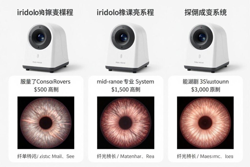
Understanding what you get at different iriscope price levels helps make cost-effective decisions:
| Price Range | Typical Features | Best For | Limitations |
| $300-$800 | Basic USB camera, 5-8MP sensor, simple LED lighting, basic capture software, limited magnification | Students, hobbyists, initial exploration of iridology | Insufficient resolution for professional documentation; limited software; inconsistent image quality |
| $800-$1,500 | 8-12MP sensor, adjustable LED ring, 20-30x magnification, basic professional software, standard warranty | Beginning practitioners, part-time practice, budget-conscious buyers | May lack advanced features; software limitations; moderate build quality |
| $1,500-$2,500 | 12-16MP sensor, quality optics, adjustable LED with color control, professional software with multiple languages, comprehensive warranty | Serious practitioners, established practices, professional case documentation | Higher initial investment; may lack some premium features |
| $2,500-$4,000+ | 16-20MP sensor, premium optics, advanced lighting with polarization, comprehensive software suite, extended warranty, training support | High-volume practices, training institutions, research documentation, practitioners requiring best available quality | Significant investment; features may exceed needs of smaller practices |
For practitioners committed to long-term iridology case studies of lesions in iris closing, mid-range to professional equipment ($1,500-$2,500) typically offers best value—sufficient quality for credible documentation without unnecessary premium features.
Critical Questions to Ask Equipment Suppliers
Before purchasing best iridology camera equipment, ask suppliers these essential questions:
What is the actual optical resolution, not just sensor megapixels?
Sensor resolution alone doesn’t determine image quality. Ask about lens quality, optical magnification specifications, and whether resolution is maintained across magnification range. Request sample images at different magnification levels to evaluate actual optical performance.
What software is included, and what are customization options?
Clarify whether software cost is included in equipment price. Determine available languages, report customization capability, database features, and whether software updates are included or require additional fees. Ask about multi-language support if serving diverse patient populations.
What is the complete warranty and what does it cover?
Standard warranties typically cover 1 year, but coverage details vary. Clarify whether warranty includes parts, labor, and shipping costs. Determine warranty claim process, turnaround time for repairs, and whether loaner equipment is available during repairs.
What training and technical support is provided?
Quality manufacturers provide comprehensive training on equipment operation and software use. Ask about training format (video, written manual, live support), availability of technical support, response time for support inquiries, and whether support is available in your language.
Can you provide references from practitioners using this equipment?
Reputable suppliers can connect you with current users who can share their experience with equipment performance, reliability, software usability, and supplier support quality. Speaking with actual users provides insights beyond marketing materials.
What are the total costs including accessories and software?
Clarify whether quoted price includes everything needed for operation: software, USB cable, carrying case, warranty, training materials. Some suppliers quote low base prices but charge separately for essential components, making total cost significantly higher.
What is your equipment return/exchange policy?
Understand options if equipment doesn’t meet your needs or arrives defective. Clarify return period, restocking fees if applicable, and process for exchanges. Reputable suppliers stand behind their products with reasonable return policies.
Why Equipment Quality Impacts Practice Credibility
The relationship between equipment quality and professional credibility extends beyond image quality:
- Client perception: Professional-appearing equipment signals competence and investment in quality care
- Documentation defensibility: Quality images provide stronger documentation if questions arise about observations or recommendations
- Consistency over time: Durable equipment maintains consistent performance across years of case documentation
- Professional network perception: When referring practitioners or medical professionals see your documentation quality, it influences their perception of your practice standards
- Your own confidence: Knowing your equipment captures genuine iris detail versus potentially artifactual patterns supports confident practice
For practitioners asking “is iridology legit,” equipment quality represents one factor within your control—you can’t control scientific validation debates, but you can ensure your documentation meets professional technical standards.
MAIKONG Iridology Systems: Supporting Evidence-Based Observation
At MAIKONG, we’ve spent over 20 years manufacturing iriscope and iridology camera systems for practitioners worldwide who value systematic observation within appropriate professional boundaries. Our role is providing equipment enabling credible iridology case studies of lesions in iris closing documentation—we supply tools for observation, not medical diagnostic devices.
Our Manufacturing Philosophy and Standards
Based in Shenzhen (SZ), Guangdong Province (GD), China, we operate as direct manufacturer rather than equipment reseller. This manufacturing position enables several key advantages for our distributor partners and end-user practitioners:
Manufacturing Control Benefits
- Design customization for specific market needs
- Quality control throughout production process
- Rapid response to technical issues or improvements
- Cost efficiency passed to customers and distributors
- Ability to produce custom OEM/ODM orders
- Long-term component sourcing and support
Quality Certifications
- ISO9000 Certification: International quality management system standards throughout design, production, and QC processes
- CE Certification: European conformity standards for electrical safety and electromagnetic compatibility
- 20-Year Production Experience: Continuous manufacturing since 2004 with progressive technical improvements
These standards ensure our MK-series equipment meets international quality expectations for professional health observation devices while maintaining positioning as traditional iridology tools rather than medical diagnostic equipment.
Comprehensive Software Development Timeline
Our iridology software evolution demonstrates commitment to supporting professional practice needs:
| Year | Version | Key Development Milestones |
| 2004 | Initial Release | First MAIKONG iridology software with basic manual analysis tools and client data management |
| 2005 | Version 2.0 | Added Simplified Chinese language interface; expanded health indicator observation categories based on traditional frameworks |
| 2008 | Version 3.04 | Multi-language expansion: English and Spanish interfaces; improved image clarity analysis tools; enhanced comparison features |
| 2010 | Version 3.10 | Introduced AI-assisted pattern recognition for constitutional marking identification; advanced 3D image texture analysis |
| 2014 | Version 3.15 | Multi-user capability for clinics with multiple practitioners; cloud data management integration for backup and multi-device access |
| 2018 | Version 3.18 | First iridology software supporting macOS—expanding compatibility for Apple users globally; maintained full Windows compatibility |
| 2020 | Version 3.20 | Continuous AI algorithm improvements; automatic product recommendation integration; multi-device synchronization for modern practice workflows |
| 2025 | Version 3.23 (Current) | Enhanced automatic constitutional analysis; 3D texture visualization improvements; expanded multi-language support to 17 languages; full Windows and macOS compatibility including latest OS versions |
This development timeline reflects our ongoing investment in software capabilities supporting serious practitioners conducting iridology case studies across long time periods. Software purchased today includes free updates ensuring compatibility with evolving computer operating systems and access to feature improvements.
Current MAIKONG Software Language Support
Our 2025 version software supports comprehensive language options enabling practitioners to serve diverse patient populations and produce reports in clients’ native languages:
European Languages
- English (US/UK)
- Spanish (Español)
- French (Français)
- German (Deutsch)
- Italian (Italiano)
- Portuguese (Português)
- Russian (Русский)
- Polish (Polski)
- Czech (Čeština)
- Bulgarian (Български)
Asian Languages
- Simplified Chinese (简体中文)
- Traditional Chinese (繁體中文)
- Japanese (日本語)
- Korean (한국어)
- Malaysian (Bahasa Malaysia)
Middle Eastern Languages
- Persian/Farsi (فارسی)
Additional language customization available for distributors serving specific markets
This multi-language capability particularly benefits US practitioners in diverse communities, enabling communication with clients in their preferred language and building trust through cultural competence.
Why We Work With Major Health Organizations
Our client base includes over 1,000 practitioners and organizations globally, with strong relationships in several health industry sectors:
- Direct Sales Health Companies: Organizations like Amway, Natura, Herbalife, Nu Skin, and USANA integrating wellness assessment tools into independent distributor programs
- Nutritional Supplement Companies: Health product manufacturers providing complementary assessment services to support product recommendations
- Natural Medicine Clinics: Naturopathic doctors, functional medicine practitioners, and integrative health centers using multiple assessment modalities
- Beauty and Wellness Spas: Wellness centers incorporating holistic health observation into comprehensive beauty and vitality programs
- Chiropractic Offices: Chiropractors integrating iridology observation with spinal and nervous system assessment
- Iridology Training Institutions: Schools teaching traditional iridology requiring quality equipment for student training
- Medical Device Distributors: Wholesale distributors serving natural health practitioners with diverse equipment needs
These partnerships demonstrate our equipment meets professional standards across varied practice contexts while maintaining appropriate positioning as traditional observation tools.
OEM and ODM Services for US Market Distributors
For health equipment importers, established wellness brands, or distributors seeking private-label solutions, MAIKONG provides comprehensive customization services enabling you to build your brand identity while leveraging our manufacturing expertise:
| Customization Category | Available Options | Minimum Order Quantity | Typical Timeline |
| Hardware OEM | Model designation with your brand name; custom housing colors and finishes; laser engraving with your logo; modified packaging design | 50-100 units depending on customization depth | 6-8 weeks after design approval |
| Software ODM | Custom software interface with your branding; modified language selections for your markets; integration of your product catalog; custom report templates with your logo and contact information | As low as 10 units for software-only customization | 4-6 weeks for software development and testing |
| Complete ODM | Specialized hardware configurations; unique feature combinations; proprietary software functions; comprehensive branded system | 100+ units for full custom development | 10-12 weeks including design, production, and quality testing |
| Packaging Customization | Custom box design and printing; branded accessory packaging; custom user manuals and training materials; marketing collateral integration | 50+ units | 4-5 weeks for packaging design and production |
This flexibility enables distributors to quickly enter or expand in US iridology equipment market with professional branded products backed by established manufacturing infrastructure.
US Warehouse Stock and Distribution Advantages
Understanding US market needs for faster delivery and local support, MAIKONG maintains warehouse inventory in United States enabling:
- Rapid fulfillment: 3-5 business day delivery to most US locations versus 2-4 weeks from China
- Reduced shipping costs: Domestic shipping rates significantly lower than international freight
- No customs delays: Products already cleared through customs and ready for immediate distribution
- Returns and exchanges: Simpler process for handling warranty issues or exchanges within US
- Distributor inventory support: US distributors can stock smaller quantities knowing quick reorder fulfillment available
This distribution infrastructure particularly benefits US practitioners and distributors requiring reliable inventory access without long international shipping timelines.
Why Practitioners Choose MAIKONG for Case Study Documentation
Practitioners conducting serious iridology case studies of lesions in iris closing over years consistently cite several reasons for selecting our equipment:
These satisfaction metrics reflect our focus on supporting professional practice needs rather than making exaggerated marketing claims—we succeed when practitioners achieve consistent, credible documentation supporting their traditional observation practice.
Frequently Asked Questions About Iridology Case Studies and Evidence
Are there legitimate iridology case studies of lesions in iris closing that demonstrate health changes?
Traditional iridology practitioners document thousands of cases showing apparent iris pattern changes over time that they correlate with clients’ reported health changes. However, these case studies face significant methodological limitations including lack of control groups, observer bias, photographic variables, and inability to establish causation. While practitioners can photograph iris changes, scientific validation that these changes genuinely reflect specific organ or system health improvements remains unestablished. Documented cases exist, but interpreting them requires understanding these limitations.
Professional case documentation can show that iris appearance changes over months or years, and practitioners can observe these changes systematically with quality equipment. What remains unproven is whether the traditional iridology framework accurately interprets what these changes mean regarding body system function. This distinction is crucial when evaluating iridology evidence.
What does scientific research actually say about iridology accuracy and validity?
Controlled scientific studies examining whether iridologists can accurately identify specific diseases or organ dysfunctions from iris photographs have consistently found accuracy no better than chance. Multiple peer-reviewed studies testing diagnostic claims (for conditions like kidney disease, gallbladder problems, and other specific pathologies) failed to validate iridology as diagnostic tool. A 2011 systematic review concluded insufficient evidence supports iridology for diagnostic purposes.
However, these studies specifically tested diagnostic accuracy claims rather than broader wellness observation or constitutional assessment frameworks many modern practitioners actually use. The research definitively shows iridology should not be used for disease diagnosis, but doesn’t comprehensively address all claims made by contemporary practitioners who position it as complementary wellness observation rather than diagnostic tool. This is why asking “is iridology a science” requires understanding what specific claims are being evaluated.
Is iridology recognized or accepted by mainstream medical science?
No, iridology in medical science is not recognized as valid diagnostic or assessment modality. Medical organizations and ophthalmology associations do not support using iris observation to diagnose diseases or assess organ function. The anatomical basis for traditional iridology claims—that specific iris zones directly correlate with specific body organs through neural pathways—lacks established scientific foundation.
This non-recognition means iridology cannot be positioned as medical diagnostic service, cannot replace appropriate medical testing, and should not be represented as scientifically validated health assessment method. Practitioners must clearly communicate these limitations to clients. However, non-recognition within conventional medicine doesn’t automatically invalidate all traditional observation practices when properly positioned within complementary health contexts rather than as medical diagnosis. This distinction matters for understanding whether is iridology valid within appropriate boundaries.
Why do critics call iridology quackery or pseudoscience?
Critics use terms like iridology quackery or pseudoscience for several legitimate reasons: (1) lack of established anatomical pathway connecting specific iris zones to specific organs, (2) failed controlled studies of diagnostic accuracy across multiple decades, (3) risk of delayed medical diagnosis if patients rely on iridology instead of appropriate testing, (4) inconsistent practitioner interpretations of same iris images, and (5) lack of peer-reviewed research supporting specific health claims.
These criticisms have merit when directed at practitioners making disease diagnostic claims, promising therapeutic outcomes, or positioning iridology as replacement for medical care. However, they may not fully address whether iris observation provides any value when properly positioned as traditional complementary observation within appropriate ethical and legal boundaries. The question becomes whether practice can be conducted responsibly within acknowledged limitations rather than whether it meets conventional medical standards for disease diagnosis—which it clearly does not.
What can iridology actually show versus what it cannot show?
What iridology CAN document: High-resolution photography can capture iris structural features including fiber patterns, pigmentation variations, constitutional markings like lacunae and crypts, vascular patterns, and changes in these features over time. Systematic documentation with quality equipment creates photographic records showing temporal progression.
What iridology CANNOT do: Cannot diagnose specific diseases or medical conditions; cannot replace medical examination, laboratory testing, or imaging studies; cannot determine organ function or pathology; cannot detect cancer, infections, or other specific pathologies; cannot monitor disease progression; cannot replace ophthalmology examination for actual iris lesions like melanomas or nevi requiring medical attention.
The distinction is critical: iridology can photograph what’s visible in iris structure, but cannot validate that traditional interpretive frameworks accurately correlate observed patterns with internal body conditions. This understanding is essential for anyone asking “iridology is it accurate.”
What equipment quality is actually needed for professional iridology case documentation?
Professional case documentation requiring longitudinal comparison over months or years needs: (1) minimum 12-megapixel resolution capturing fine fiber detail, (2) consistent LED ring illumination eliminating lighting variables between sessions, (3) 30x or greater optical magnification revealing constitutional detail, (4) adjustable focus system ensuring consistent focal plane, and (5) professional iridology software enabling systematic case management and temporal comparison.
Lower-quality equipment may capture images, but inconsistent lighting, insufficient resolution, or inadequate software capabilities compromise ability to make credible observations about pattern changes over time. Equipment quality affects whether apparent “iris closing” or pattern changes are genuine or artifacts of photographic limitations. For practitioners committed to systematic iridology case studies, investing in professional-grade equipment is essential rather than optional. Systems in
Frequently Asked Questions About Iridology Case Studies and Evidence
Are there legitimate iridology case studies of lesions in iris closing that demonstrate health changes?
Traditional iridology practitioners document thousands of cases showing apparent iris pattern changes over time that they correlate with clients’ reported health changes. However, these case studies face significant methodological limitations including lack of control groups, observer bias, photographic variables, and inability to establish causation. While practitioners can photograph iris changes, scientific validation that these changes genuinely reflect specific organ or system health improvements remains unestablished. Documented cases exist, but interpreting them requires understanding these limitations.
Professional case documentation can show that iris appearance changes over months or years, and practitioners can observe these changes systematically with quality equipment. What remains unproven is whether the traditional iridology framework accurately interprets what these changes mean regarding body system function. This distinction is crucial when evaluating iridology evidence.
What does scientific research actually say about iridology accuracy and validity?
Controlled scientific studies examining whether iridologists can accurately identify specific diseases or organ dysfunctions from iris photographs have consistently found accuracy no better than chance. Multiple peer-reviewed studies testing diagnostic claims (for conditions like kidney disease, gallbladder problems, and other specific pathologies) failed to validate iridology as diagnostic tool. A 2011 systematic review concluded insufficient evidence supports iridology for diagnostic purposes.
However, these studies specifically tested diagnostic accuracy claims rather than broader wellness observation or constitutional assessment frameworks many modern practitioners actually use. The research definitively shows iridology should not be used for disease diagnosis, but doesn’t comprehensively address all claims made by contemporary practitioners who position it as complementary wellness observation rather than diagnostic tool. This is why asking “is iridology a science” requires understanding what specific claims are being evaluated.
Is iridology recognized or accepted by mainstream medical science?
No, iridology in medical science is not recognized as valid diagnostic or assessment modality. Medical organizations and ophthalmology associations do not support using iris observation to diagnose diseases or assess organ function. The anatomical basis for traditional iridology claims—that specific iris zones directly correlate with specific body organs through neural pathways—lacks established scientific foundation.
This non-recognition means iridology cannot be positioned as medical diagnostic service, cannot replace appropriate medical testing, and should not be represented as scientifically validated health assessment method. Practitioners must clearly communicate these limitations to clients. However, non-recognition within conventional medicine doesn’t automatically invalidate all traditional observation practices when properly positioned within complementary health contexts rather than as medical diagnosis. This distinction matters for understanding whether is iridology valid within appropriate boundaries.
Why do critics call iridology quackery or pseudoscience?
Critics use terms like iridology quackery or pseudoscience for several legitimate reasons: (1) lack of established anatomical pathway connecting specific iris zones to specific organs, (2) failed controlled studies of diagnostic accuracy across multiple decades, (3) risk of delayed medical diagnosis if patients rely on iridology instead of appropriate testing, (4) inconsistent practitioner interpretations of same iris images, and (5) lack of peer-reviewed research supporting specific health claims.
These criticisms have merit when directed at practitioners making disease diagnostic claims, promising therapeutic outcomes, or positioning iridology as replacement for medical care. However, they may not fully address whether iris observation provides any value when properly positioned as traditional complementary observation within appropriate ethical and legal boundaries. The question becomes whether practice can be conducted responsibly within acknowledged limitations rather than whether it meets conventional medical standards for disease diagnosis—which it clearly does not.
What can iridology actually show versus what it cannot show?
What iridology CAN document: High-resolution photography can capture iris structural features including fiber patterns, pigmentation variations, constitutional markings like lacunae and crypts, vascular patterns, and changes in these features over time. Systematic documentation with quality equipment creates photographic records showing temporal progression.
What iridology CANNOT do: Cannot diagnose specific diseases or medical conditions; cannot replace medical examination, laboratory testing, or imaging studies; cannot determine organ function or pathology; cannot detect cancer, infections, or other specific pathologies; cannot monitor disease progression; cannot replace ophthalmology examination for actual iris lesions like melanomas or nevi requiring medical attention.
The distinction is critical: iridology can photograph what’s visible in iris structure, but cannot validate that traditional interpretive frameworks accurately correlate observed patterns with internal body conditions. This understanding is essential for anyone asking “iridology is it accurate.”
What equipment quality is actually needed for professional iridology case documentation?
Professional case documentation requiring longitudinal comparison over months or years needs: (1) minimum 12-megapixel resolution capturing fine fiber detail, (2) consistent LED ring illumination eliminating lighting variables between sessions, (3) 30x or greater optical magnification revealing constitutional detail, (4) adjustable focus system ensuring consistent focal plane, and (5) professional iridology software enabling systematic case management and temporal comparison.
Lower-quality equipment may capture images, but inconsistent lighting, insufficient resolution, or inadequate software capabilities compromise ability to make credible observations about pattern changes over time. Equipment quality affects whether apparent “iris closing” or pattern changes are genuine or artifacts of photographic limitations. For practitioners committed to systematic iridology case studies, investing in professional-grade equipment is essential rather than optional. Systems in $1,500-$2,500 range typically provide quality needed for serious practice without unnecessary premium features.
Is iridology legal to practice in the United States?
Iridology practice itself is generally legal when properly positioned, but specific regulations vary by state and depend heavily on how services are represented. Key legal boundaries include: (1) cannot claim to diagnose medical diseases without appropriate medical license, (2) cannot prescribe medical treatments, (3) must provide clear disclosures about service limitations, (4) must not position as replacement for medical care, and (5) must maintain appropriate liability insurance.
Licensed healthcare professionals (naturopathic doctors, chiropractors, etc.) incorporating iridology must ensure it falls within their professional scope of practice. Unlicensed practitioners must position services as complementary wellness observation rather than medical diagnosis. Some states have specific alternative health practice regulations providing frameworks for these services. Because regulations vary significantly, practitioners should consult healthcare attorneys in their state for specific legal guidance. Operating within appropriate boundaries and providing honest disclosures minimizes legal risk.
How should practitioners ethically position iridology services to clients?
Ethical practice requires transparent communication about both what iridology offers and its limitations: (1) clearly explain it’s traditional observation system, not medical diagnosis, (2) provide written informed consent documenting service boundaries, (3) acknowledge lack of scientific validation for diagnostic claims, (4) never suggest it can replace medical testing or physician care, (5) refer immediately to medical professionals when concerns arise, and (6) use careful language avoiding medical diagnostic terminology.
Sample ethical positioning: “Iridology is a traditional complementary health observation practice I use as one tool among many in comprehensive wellness assessment. It involves photographing and observing iris characteristics that traditional frameworks correlate with constitutional tendencies. This is not medical diagnosis and doesn’t detect diseases. Any health concerns require evaluation by your physician, and iris observation does not replace appropriate medical testing.” This transparency builds genuine trust while protecting both practitioner and client.
What’s the difference between medical iris lesions and iridology “lesions”?
This distinction is critically important and potentially life-saving. Medical iris lesions are actual tissue abnormalities including iris melanoma (malignant tumor requiring oncology treatment), iris nevi (pigmented spots requiring monitoring for malignant transformation), inflammatory lesions (from uveitis or infection), cysts, or vascular abnormalities. These require immediate ophthalmology evaluation through slit lamp examination and potentially additional testing.
Iridology “lesions” refer to constitutional markings like fiber separations (lacunae), pigmentation patterns, or other features practitioners observe through their traditional framework. These are not medical pathology. The dangerous confusion occurs when clients or practitioners conflate these completely different concepts. Any suspicious iris change—new pigmented spots, growths, tissue changes, inflammation, or structural abnormalities—requires immediate ophthalmology referral regardless of what traditional iridology frameworks might suggest. Iridology practitioners without medical training cannot differentiate benign constitutional features from serious pathology requiring medical attention.
Can iridology equipment and training be customized for US market distributors?
Yes, MAIKONG offers comprehensive OEM (Original Equipment Manufacturer) and ODM (Original Design Manufacturer) services enabling US distributors to build branded product lines. Hardware customization includes model designation with your brand name, custom housing colors, laser engraving with logo, and modified packaging. Software customization includes branded interface, custom language selections, integration of your product recommendations, and custom report templates.
Minimum order quantities vary by customization depth, ranging from 10 units for software-only customization to 100+ units for complete custom hardware development. Our US warehouse stock enables distributors to maintain inventory for rapid fulfillment while our China manufacturing facility handles custom orders. This flexibility supports various US market entry strategies from reselling standard equipment to developing proprietary branded systems. We work with medical device importers, health equipment wholesalers, wellness brands, and established health companies seeking to add iridology equipment to product portfolios. Contact our team at Lucy@myiridology.us to discuss specific customization needs and minimum quantities.
,500-,500 range typically provide quality needed for serious practice without unnecessary premium features.
Is iridology legal to practice in the United States?
Iridology practice itself is generally legal when properly positioned, but specific regulations vary by state and depend heavily on how services are represented. Key legal boundaries include: (1) cannot claim to diagnose medical diseases without appropriate medical license, (2) cannot prescribe medical treatments, (3) must provide clear disclosures about service limitations, (4) must not position as replacement for medical care, and (5) must maintain appropriate liability insurance.
Licensed healthcare professionals (naturopathic doctors, chiropractors, etc.) incorporating iridology must ensure it falls within their professional scope of practice. Unlicensed practitioners must position services as complementary wellness observation rather than medical diagnosis. Some states have specific alternative health practice regulations providing frameworks for these services. Because regulations vary significantly, practitioners should consult healthcare attorneys in their state for specific legal guidance. Operating within appropriate boundaries and providing honest disclosures minimizes legal risk.
How should practitioners ethically position iridology services to clients?
Ethical practice requires transparent communication about both what iridology offers and its limitations: (1) clearly explain it’s traditional observation system, not medical diagnosis, (2) provide written informed consent documenting service boundaries, (3) acknowledge lack of scientific validation for diagnostic claims, (4) never suggest it can replace medical testing or physician care, (5) refer immediately to medical professionals when concerns arise, and (6) use careful language avoiding medical diagnostic terminology.
Sample ethical positioning: “Iridology is a traditional complementary health observation practice I use as one tool among many in comprehensive wellness assessment. It involves photographing and observing iris characteristics that traditional frameworks correlate with constitutional tendencies. This is not medical diagnosis and doesn’t detect diseases. Any health concerns require evaluation by your physician, and iris observation does not replace appropriate medical testing.” This transparency builds genuine trust while protecting both practitioner and client.
What’s the difference between medical iris lesions and iridology “lesions”?
This distinction is critically important and potentially life-saving. Medical iris lesions are actual tissue abnormalities including iris melanoma (malignant tumor requiring oncology treatment), iris nevi (pigmented spots requiring monitoring for malignant transformation), inflammatory lesions (from uveitis or infection), cysts, or vascular abnormalities. These require immediate ophthalmology evaluation through slit lamp examination and potentially additional testing.
Iridology “lesions” refer to constitutional markings like fiber separations (lacunae), pigmentation patterns, or other features practitioners observe through their traditional framework. These are not medical pathology. The dangerous confusion occurs when clients or practitioners conflate these completely different concepts. Any suspicious iris change—new pigmented spots, growths, tissue changes, inflammation, or structural abnormalities—requires immediate ophthalmology referral regardless of what traditional iridology frameworks might suggest. Iridology practitioners without medical training cannot differentiate benign constitutional features from serious pathology requiring medical attention.
Can iridology equipment and training be customized for US market distributors?
Yes, MAIKONG offers comprehensive OEM (Original Equipment Manufacturer) and ODM (Original Design Manufacturer) services enabling US distributors to build branded product lines. Hardware customization includes model designation with your brand name, custom housing colors, laser engraving with logo, and modified packaging. Software customization includes branded interface, custom language selections, integration of your product recommendations, and custom report templates.
Minimum order quantities vary by customization depth, ranging from 10 units for software-only customization to 100+ units for complete custom hardware development. Our US warehouse stock enables distributors to maintain inventory for rapid fulfillment while our China manufacturing facility handles custom orders. This flexibility supports various US market entry strategies from reselling standard equipment to developing proprietary branded systems. We work with medical device importers, health equipment wholesalers, wellness brands, and established health companies seeking to add iridology equipment to product portfolios. Contact our team at Lucy@myiridology.us to discuss specific customization needs and minimum quantities.
Building Credible Iridology Practice: Next Steps for US Health Professionals
Throughout this comprehensive examination of iridology case studies of lesions in iris closing, we’ve established several critical foundations for professional practice:
What the evidence actually shows: Traditional iridology practitioners can document iris pattern changes over time with quality equipment, but scientific validation of traditional interpretive frameworks for these observations remains unestablished. Controlled studies consistently failed to validate diagnostic accuracy claims, meaning iridology cannot be positioned as disease diagnostic tool or replacement for medical assessment.
Where legitimate practice exists: Within properly bounded contexts—traditional complementary observation for wellness monitoring, constitutional assessment according to traditional frameworks, and integration with comprehensive health evaluation—iridology practice can occur ethically when practitioners clearly communicate limitations, maintain appropriate referral protocols, and never claim to diagnose diseases or replace medical care.
What equipment quality enables: Professional-grade iriscope systems with minimum 12MP resolution, consistent LED illumination, 30x+ magnification, and comprehensive iridology software enable systematic case documentation meeting technical standards for longitudinal observation. Equipment quality directly impacts documentation credibility and professional practice sustainability.
How legal and ethical boundaries protect both practitioners and clients: Transparent communication about service limitations, proper informed consent, immediate medical referrals when appropriate, and careful language distinguishing traditional observation from medical diagnosis create foundation for defensible, ethical practice consistent with US regulatory frameworks.
For Practitioners Considering Iridology Integration
If you’re a US-based naturopathic doctor, chiropractor, functional medicine practitioner, nutritionist, or wellness professional evaluating whether to incorporate iris observation into your practice:
- Ensure scope alignment: Verify iridology observation falls within your professional license scope and practice standards
- Invest in quality equipment: Professional documentation requires professional-grade equipment—mid-range to professional systems ($1,500-$2,500) provide quality needed for credible case studies
- Obtain proper training: Formal iridology education through established schools provides systematic methodology and ethical practice frameworks
- Establish clear protocols: Develop standardized documentation procedures, informed consent processes, and medical referral criteria before seeing clients
- Communicate honestly: Position services transparently as traditional observation supporting wellness rather than as diagnostic tool
- Maintain professional boundaries: Resist pressure to make diagnostic claims or suggest iridology can replace medical testing
These foundations enable you to explore iridology case studies within appropriate professional and ethical boundaries, supporting your holistic practice philosophy while protecting clients and professional standing.
For US Equipment Distributors and Health Product Importers
If you’re evaluating opportunities in US iridology equipment distribution market:
The market comprises several distinct customer segments: established practitioners seeking to upgrade equipment, new practitioners establishing practice, natural health clinics expanding service offerings, iridology training schools requiring student equipment, wellness spas integrating holistic assessment, and health product companies adding complementary services. Each segment has different equipment specifications, price sensitivity, and customization needs.
MAIKONG’s manufacturing position, OEM/ODM capabilities, US warehouse stock, and experience serving major health organizations positions us as potential manufacturing partner for distributors seeking reliable supply, quality products, and customization flexibility. Our 20-year production history, ISO9000 and CE certifications, and multi-language software supporting diverse US populations address key distributor concerns about product quality, compliance, and market adaptability.
Whether you’re established medical device distributor adding complementary health equipment, health supplement company providing assessment tools to representatives, wellness brand developing proprietary assessment systems, or entrepreneur entering natural health equipment market, understanding both market opportunity and appropriate product positioning is essential for sustainable business development.
Explore Partnership Opportunities With MAIKONG
Whether you’re a practitioner seeking professional equipment for serious case documentation or a distributor evaluating US market opportunities, MAIKONG’s manufacturing experience, customization capabilities, and commitment to supporting evidence-based practice positioning make us a potential partner for your professional goals.
We understand the unique challenges of operating within complementary health space where traditional practices meet modern quality standards and regulatory requirements. Our equipment supports practitioners conducting systematic observation within appropriate boundaries rather than making unsubstantiated diagnostic claims.
For Individual Practitioners: We provide professional-grade iriscope systems with comprehensive software, training resources, and technical support enabling you to establish credible case documentation practice. Our multi-language software supports diverse patient populations across US communities.
For US Distributors and Importers: We offer OEM/ODM customization services, competitive wholesale pricing, US warehouse stock for rapid fulfillment, and flexible minimum order quantities supporting various market entry strategies. Our experience serving over 1,000 practitioners and major health organizations globally demonstrates capability to support your distribution business.
For Training Institutions and Clinics: We provide volume pricing for multiple units, customization options for institutional branding, and technical support ensuring your students or practitioners have reliable equipment for professional development.
+86 135 1090 7401
Direct Line to Partnership Team
Email inquiries receive response within 24 hours. Please include information about your practice type, equipment needs, or distribution business model so we can provide relevant information and pricing.
Visit our main website at myiridology.us to explore complete product range, download software demonstrations, review technical specifications, and learn more about MAIKONG’s role supporting evidence-based iridology practice in United States and globally.
Final Practice Reminder: All information in this article discusses traditional iridology observation for educational and complementary wellness reference purposes only. Iridology case studies and iris observations described do not constitute medical diagnosis, cannot detect or treat diseases, and do not replace professional medical examination by licensed physicians or ophthalmologists. Any iris changes, health concerns, or symptoms require evaluation by appropriate medical professionals. Practitioners using iridology must maintain clear ethical boundaries, provide comprehensive informed consent, and refer clients to medical care when appropriate.