
Ciliary Zone Iridology: How to Read, Compare, and Use an Iridology Chart
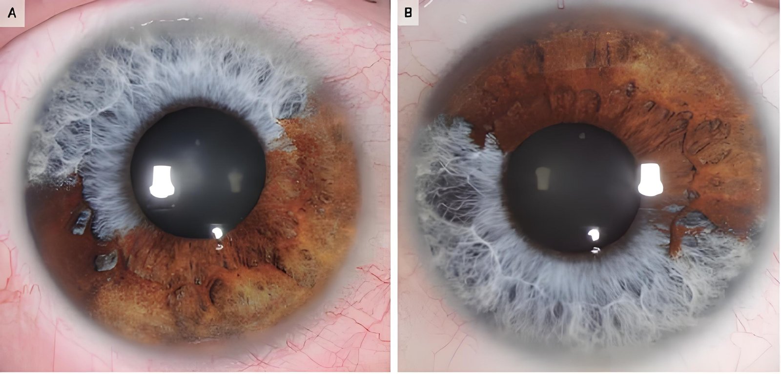
In traditional iridology, the ciliary zone represents the broad outer region of the iris located between the autonomic nerve wreath and the outer ciliary border. Practitioners read this circular band using specialized reference charts that divide the iris stroma into concentric rings and radial sectors corresponding to peripheral organs, circulation, and structural systems. Rather than delivering a medical diagnosis, a ciliary zone iridology chart serves as an educational reference map that helps observers evaluate structural density, pigment variations, and constitutional patterns.
Selecting the right chart approach depends on whether the material is needed for client consultations, personal study, classroom training, or digital image overlays within specialized analysis software. Proper lighting, calibrated optical capture, and bilateral eye comparison decide how accurately an observer can align physical iris landmarks with printed or digital coordinate maps.
What Is Ciliary Zone Iridology?
Traditional iris evaluation divides the visible stroma into two primary physiological areas: the inner pupillary zone and the outer ciliary zone. The boundary separating these two regions is the collarette, also known in classical iridology as the autonomic nerve wreath. In the study of ciliary zone iridology, observers focus on the expansive outer band that extends from this raised zigzag line all the way to the limbus or scleral margin.
While the narrow pupillary zone primarily reflects the stomach and intestinal tract in reflex models, the ciliary zone encompasses the vast majority of systemic representations. Reflexology charts place major organ systems within this zone, including the liver, kidneys, lungs, endocrine glands, musculoskeletal frame, lymphatic networks, and outer skin perimeter. Reading this area requires understanding how classical cartographers transferred three-dimensional biological tissue onto flat, circular topological maps.
Ciliary zone maps are educational references designed to reflect systemic vitality and constitutional predispositions, rather than diagnostic tools for detecting specific pathological illnesses.
Practitioners utilize an iridology chart and the body reflex model to note variations in fiber density, coloration, openings, and shading. These traditional observations provide insight into inherited constitutional tendencies and functional stress points across the body’s interrelated physical networks.
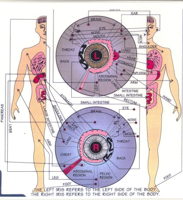
The ciliary zone extends outward from the collarette line to the outer iris perimeter.
Where the Ciliary Zone Appears on an Iridology Chart
Locating the ciliary zone on a physical or digital iris map requires identifying the foundational concentric rings established by European and American charting systems. Classical charts divide the entire iris into seven concentric rings, starting at the pupil edge and moving outward toward the sclera. Rings three through seven sit entirely within the ciliary zone, spanning the humoral circulation zone, the parenchymal organ zone, the lymphatic belt, the motor-sensory ring, and the skin margin.
In addition to concentric rings, an iridology chart map incorporates clock-face coordinates divided into sixty minute marks or twelve primary hour sections. Radial sectors project outward from the central collarette toward the perimeter, positioning organs in predictable topological sectors. This dual-axis coordinate system enables practitioners to locate reflexive areas with high spatial precision.
Inner Ciliary Margin
Directly borders the collarette, housing reflex points for deep vascular and autonomic nervous balance.
Middle Ciliary Zone
Contains the reflex locations for major parenchymal organs such as the kidneys, liver, lungs, and reproductive tract.
Outer Ciliary Perimeter
Represents the lymphatic circulation, circulatory beds, and the scurf rim representing dermal elimination.
When studying an iridology chart of eye reflexology, analysts observe how the fibers radiate across these zones like spokes on a bicycle wheel. Disruptions, bends, pigment deposits, or gaps along these radial fibers indicate specific areas of constitutional interest according to historical iridology models.
Right-Eye and Left-Eye Chart Differences
Because human anatomy exhibits distinct bilateral asymmetry, iridology charts are strictly divided between the right and left iris. Single organs situated primarily on one side of the torso reflect exclusively in the corresponding eye chart. Comparing a iridology chart right eye with a left eye iridology chart prevents misidentifying crucial anatomical sectors during visual evaluation.
In a standard layout, the right eye chart maps the liver, gallbladder, ascending colon, right kidney, and right lung. Conversely, the left eye map displays the spleen, descending colon, sigmoid flexure, left kidney, and heart reflex sectors. The practitioner must also verify the nasal and temporal orientations, as the medial aspect of each eye borders the nose while the temporal aspect faces the ear.
Understand Clock Positions
chart is divided into 12-hour clock layout, and each clock position corresponds to specific organ or system. example, 1 o’clock typically represents head and brain, while 6 o’clock could represent kidneys.
I usually start with outer rim (closer to edge iridology) and work my way inward, different layers Iridology reflect deeper aspects body.
Here’s general breakdown that I rely on understande 12-hour clock positions:
| Clock Position | Left Eye (Reflects Left Side Body) | Right Eye (Reflects Right Side Body) |
|---|---|---|
| 1 o’clock – 2 o’clock | Left Neck: Reproductive organs (uterus/ovaries/testes) | Right Face: Reproductive organs (uterus/ovaries/testes) |
| 2 o’clock – 3 o’clock | Left Lung: Respiratory health | Right Throat: Health joints (knees, hips, elbows, shoulders) |
| 3 o’clock – 4 o’clock | Left Thorax: Spine health, flexibility, alignment | Right Upper Back: Spine health, flexibility, alignment |
| 4 o’clock – 5 o’clock | Left Upper Abdomen: Bladder, urinary system | Right Bladder: Reflects bladder and left side urinary system |
| 5 o’clock – 6 o’clock | Left Pelvic Area: Colon, small intestine, digestive health | Right Pelvic Area: Colon, small intestine, digestive health |
| 6 o’clock – 7 o’clock | Left Lower Abdomen: Right kidney (filtration, detox) | Right Lower Abdomen: Left kidney (filtration, detox) |
| 7 o’clock – 8 o’clock | Left Upper Abdomen: Stomach, digestive organs | Right Upper Abdomen: Stomach, digestive organs |
| 8 o’clock – 9 o’clock | Left Thorax: Liver, detoxification, bile production | Right Thorax: Liver, detoxification, bile production |
| 9 o’clock – 10 o’clock | Left Lung: Cardiovascular health, circulation | Right Lung: Cardiovascular health, circulation |
| 10 o’clock – 11 o’clock | Left Neck: Health joints (knees, hips, elbows) | Right Neck: Health joints (knees, hips, elbows) |
| 11 o’clock – 12 o’clock | Left Brain: Cognitive function, mental health | Right Brain: Cognitive function, mental health |
| 12 o’clock – 1 o’clock | Left Brain: Cognitive function, mental health | Right Brain: Cognitive function, mental health |
A practitioner using an iridology eye chart left or inspecting an iridology chart right chart must place both maps adjacent to each other for full-body balance assessments. Utilizing an iridology chart side by side format simplifies comparative analysis between paired organs. Even when an older textbook contains a mislabeled iridology chart left eyte illustration, cross-referencing nasal landmarks keeps the reading geometrically accurate.
Common Ciliary Zone Iridology Chart Formats
Educators and wellness analysts utilize diverse physical and digital formats to study the ciliary zone. Depending on clinical workflows, practitioners alternate between wall-mounted posters, laminated desk references, pocket-sized cards, and digital transparent graphic overlays. Each medium serves distinct educational and visual purposes within a professional wellness setting.
Selecting an appropriate layout requires evaluating how the chart will be referenced during client sessions or student instruction. A large format facilitates patient education from across an exam table, while compact desktop sheets permit rapid technical cross-referencing during magnification reviews. Digital formats allow software systems to scale transparency grids directly over high-definition iris photography.
Large Wall Maps
Ideal for lecture halls, clinical treatment rooms, and educational presentations where viewers observe whole-body reflex relationships from a distance.
Desk Study Sheets
Laminated reference sheets placed beside the microscope or computer display, offering high durability and rapid sector identification during consultations.
Digital Image Overlays
Calibrated transparent image templates built into specialized software to project clock rings and organ boundaries directly over live digital captures.
Regional variations also influence chart styling across different parts of the world. While American reflexology often relies on color-coded sectoral maps, an eye chart iridology resource from Europe may focus heavily on constitutional iris types and structural fiber deviations. An analyst reviewing an iridology chart uk edition will observe German-inspired constitutional classifications alongside traditional sector divisions, contrasting with regional publications like an iridology chart in idaho falls that follow classic Jensenian holistic mapping.
Printable, Desk, and Wall Chart Variations
When selecting physical documentation, practitioners evaluate the functional differences between downloadable printable files and professional commercial charts. A downloadable iridology chart printable PDF provides rapid access for classroom handouts, student field work, and client take-home summaries. However, professional clinical settings often demand heavy-stock laminated products designed to resist ambient moisture and daily clinical wear.
Display size substantially affects visual clarity during complex multi-zone analysis. An integrated iridology wall chart provides comprehensive anatomical typography that remains readable across a clinic room, making it an excellent centerpiece for explaining constitutional concepts. Conversely, an integrated iridology desk chart or an integrated iridology iris desk chart prioritizes compact utility, positioning intricate minute coordinates within immediate view of the practitioner’s focal field.
Full-Color Charts
- Clear visual separation of organ zones using distinct pastel or saturated color bands.
- Enhanced client communication during face-to-face holistic reviews.
- Clear demarcation of lymphatic, respiratory, and digestive perimeters.
Monochrome & Black-and-White
- High contrast typography suitable for standard office printing and photocopying.
- Uncluttered background that keeps focus strictly on structural fiber direction.
- Clean lines favored for precise student marking and manual annotation.
Working with an iridology chart black and white format offers distinct diagnostic advantages when evaluating stroma architecture. Removing vibrant color fills allows the practitioner to concentrate entirely on lacuna borders, nerve rings, radii solaris, and vascular variations without visual distraction.
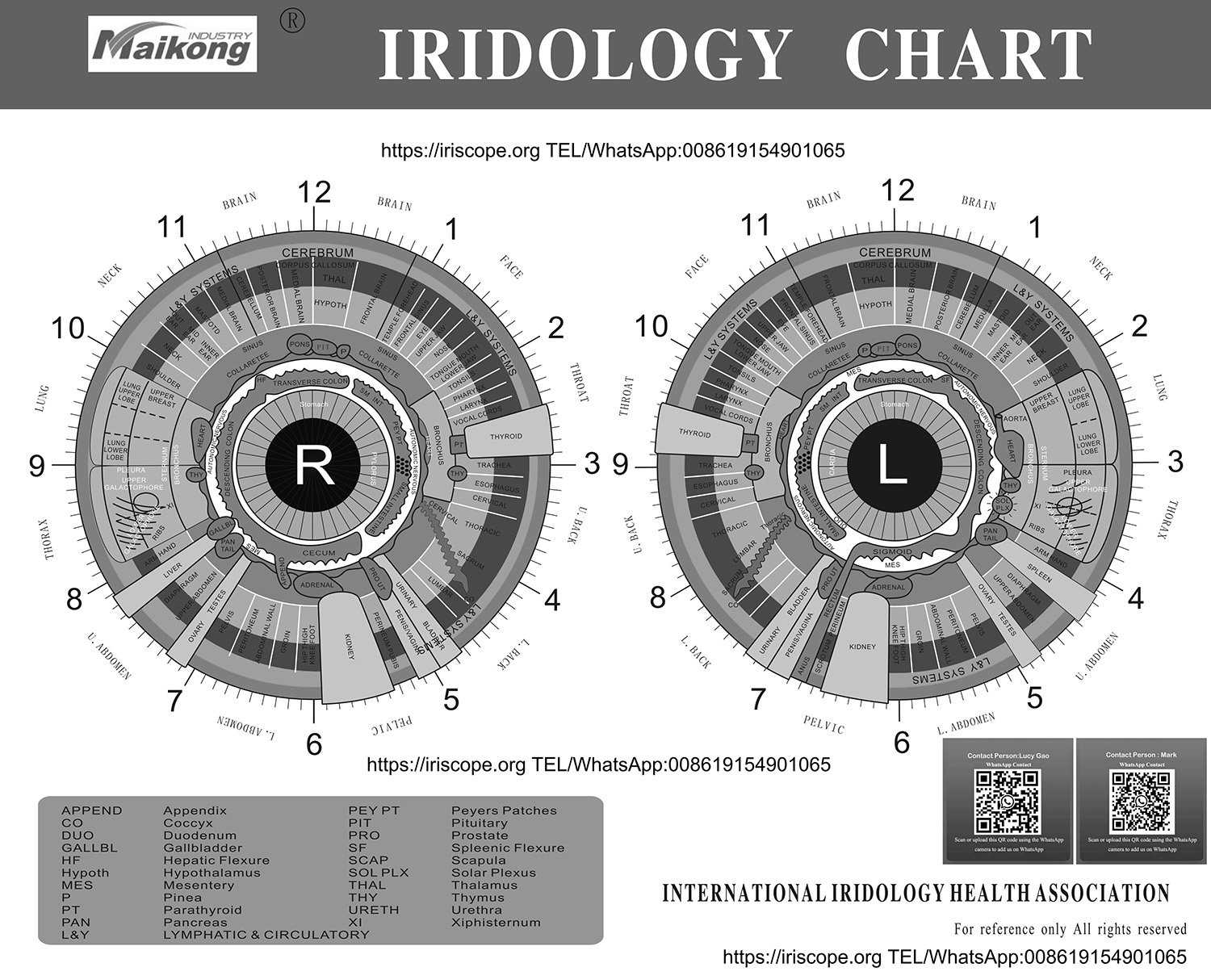
Monochrome charts provide high contrast for studying fiber alignment and structural crypts.
Integrated and Historical Chart Traditions
Modern iris evaluation builds upon over a century of empirical charting across Europe, North America, and Australia. In the United States, the pioneering work of Dr. Bernard Jensen popularized reflexive topography through the widely recognized Bernard Jensen iridology chart. This traditional paradigm links concentric ciliary rings directly to nutritional transit, bowel reflexes, and systemic vitality levels.
Subsequent researchers refined these layouts to reflect advancing anatomical understanding and constitutional typing. Significant contributions include the dr mary reed gates iridology chart, known for its focus on gland reflexes and endocrine pathways, and the detailed clinical layout of dr micki jones iridology charts. Contemporary European naturopathy frequently references an iridiagnosis iridology chart based on the historical foundations of Ignatz von Peczely and Nils Liljequist, as well as the mid-century revisions by Peter Johannes Thiel and the classic iridology chart hensen lineage.
| Tradition & Cartographer | Primary Methodological Focus | Ciliary Zone Presentation | Recommended Application |
| Jensenian Charting | Nutritional and bowel reflexology | Seven concentric rings with detailed gastrointestinal reflex arcs | Holistic nutrition, lifestyle education, and naturopathy clinics |
| Integrated Iridology | Modern constitutional synthesis | Combined genetic collarette topography and structural grading | Advanced clinical practice, pathology correlation, academic study |
| Holistic Constitutional | Constitutional summary dynamics | Broad systemic overlays linked to diathesis and pigment patterns | Integrative health assessments and patient vitality summaries |
| Herbalist Health Club | Detoxification and lymphatic filtration | Emphasis on scurf rim, kidneys, adrenals, and lymphatic stagnation | Detoxification education, botanical protocols, wellness centers |
Modern analysts frequently utilize an integrated iridology chart or an integrated iridology iris chart to harmonize classic reflexology with modern European constitutional analysis. Resources like the integrated-iridology-constitution-chart and the holistic iridology constitutional summary chart evaluate iris color variations alongside structural trabecular patterns. Practitioners focused on botanical wellness protocols often incorporate an iridology chart from drmorsesherbalhealthclub to highlight kidney filtration points and lymphatic elimination rings.
How to Read a Ciliary Zone Iridology Chart Step by Step
Interpreting the ciliary zone requires a disciplined, methodical observation workflow to avoid spatial confusion. Moving directly to isolated marks without establishing basic landmarks can lead to inaccurate sector alignment. A structured approach guarantees that the practitioner reads the correct anatomical reflex zone every time.
Understanding an iridology chart how to read manual begins by establishing correct eye identification, orientation, and magnification parameters before conducting deeper fiber analysis.
- Confirm eye orientation by identifying the caruncle, tear duct, or nasal shadow on the raw image to prevent flipping the right and left iris maps.
- Locate the collarette boundary to establish the exact dividing line between the inner pupillary zone and the outer ciliary field.
- Establish the 12 o’clock and 6 o’clock vertical axis, cross-referencing head and kidney zones to align the chart’s radial grid.
- Trace the primary concentric rings outward from the autonomic wreath toward the limbus, noting where the target fiber deviation resides.
- Read the clock sector coordinates in minutes, matching the observed stroma marking with the corresponding organ designation on an iridology chart health chart.
- Document fiber density, lacuna depths, pigment deposits, and radial furrows as educational constitutional markers without generating clinical diagnoses.
Applying this consistent procedure prevents misreading temporal markings as nasal reflexes, ensuring reliable documentation across successive patient sessions.

Following a structured reading sequence ensures accurate anatomical alignment across all iris sectors.
Evaluating Markings, Depressions, and Pigment Shifts
Fibers within the ciliary zone rarely appear as uniform, straight radial lines. In living tissue, practitioners observe an array of structural features including crypts, closed lacunae, honeycomb formations, radii solaris, and contraction furrows. In traditional terminology, a depression in iridology chart analysis represents a localized structural separation where underlying darker trabecular layers become visible through the anterior stroma leaf.
Observers often encounter dark openings described historically as an iridology chart black holes in iris pattern. Rather than indicating physical voids or acute disease states, these dark lacunae indicate areas where iris stromal fibers have separated, revealing the deeper, heavily pigmented posterior epithelium. Traditional charts classify these openings as constitutional markers of reduced structural density or areas of inherited metabolic vulnerability.
Structural depressions, crypts, and darkened trabeculae in the ciliary zone indicate inherited tissue density and constitutional tendencies, never acute infections or pathological diagnoses.
Practitioners also reference historical iridology drug and chemical charts when observing discrete exogenous pigment spots or localized topostabile color alterations. While nineteenth-century texts attributed bright yellow, rust, or iodine-toned spots to settled pharmaceuticals, modern iridology interprets these heterochromias as enzyme, liver, or metabolic markers within the reflexive organ sector.
How to Use a Chart with an Iriscope, Iridology Camera, or Software
While hand-held loupes and penlights served early practitioners, modern clinical iridology relies on digital imaging systems to capture steady, high-magnification iris photography. Utilizing a professional iriscope or dedicated iridology camera eliminates motion blur and guarantees balanced bilateral illumination across both temporal and nasal perimeters. A quality USB iridology camera connects directly to clinical computers, streaming live feeds to facilitate rapid focus adjustments.
Specialized iridology software transforms chart interpretation by superimposing transparent vector maps directly over captured pupil and ciliary zones. Practitioners can download an iridology software free download package to evaluate digital alignment tools, measure pupil decentration, and adjust chart transparency levels dynamically. Setting up a dedicated iridology camera stand ensures steady optical alignment, while keeping the CADI iridology camera driver updated guarantees seamless USB communication.
| Imaging Approach | Magnification & Clarity | Chart Integration Method | Primary Operational Advantage |
| Handheld 10X Loupe | Basic optical view | Manual side-by-side desk chart comparison | Portability for rapid field screening and classroom study |
| Standard Slit Lamp | High optical depth | External printed chart cross-referencing | Exceptional cross-sectional corneal and stromal depth viewing |
| USB Digital Iriscope | 12.0 Megapixel CCD | Direct digital software overlay with scalable transparency | Immediate client visualization, archive storage, and comparison |
Clinicians investing in the best iridology camera evaluate sensor resolution, lens coatings, and software versatility before purchasing an iridology camera for sale. Digital overlays make client communication intuitive, allowing users to see their own iris stroma mapped precisely against classical ciliary sectors on-screen.
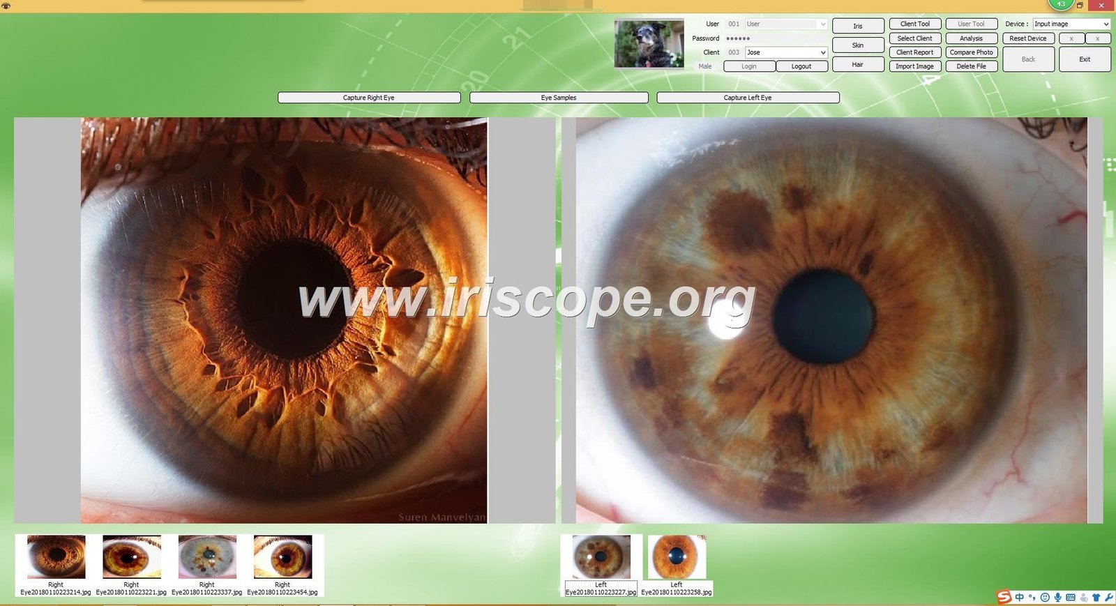
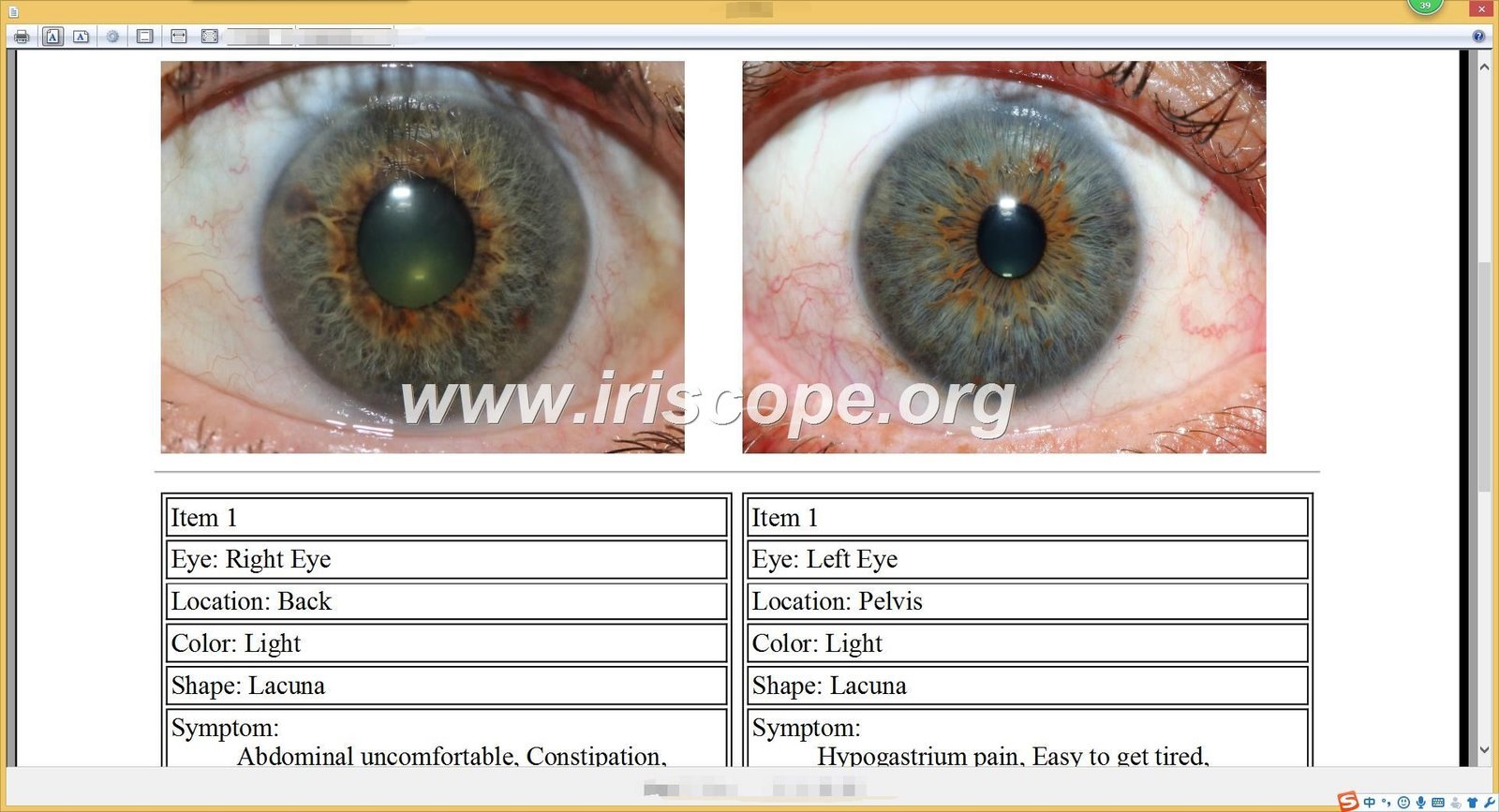
Digital software tools allow practitioners to overlay scalable ciliary zone maps directly onto patient iris images.
MAIKONG Camera and Software Specifications
MAIKONG has engineered professional optical equipment and analysis software in GD, SZ for over twenty years. Operating under comprehensive ISO9000 production guidelines, we manufacture high-precision iriscope systems designed to assist practitioners in accurate ciliary zone mapping. For commercial buyers and natural health distributors evaluating the iriscope price, our direct manufacturing capabilities ensure consistent hardware reliability and flexible customization options.
Our flagship MK-GY-9822U imaging system features custom optics and stabilized LED illumination designed specifically for stromal clarity. The technical parameters and accessories below reflect our export standard for wellness clinics and international distributors partnering with leading iridology camera manufacturers.
| Specification Parameter | Standard Factory Details |
| Brand Name | MAIKONG |
| Hardware Model | MK-9822U |
| Instrument Type | USB Iriscope / Iridology Camera |
| Sensor Resolution | 12.00 Mega Pixels High Resolution CCD |
| Image Processing | Special DSP Image Processor with OIS |
| Lens Assembly | Imported Plated Layer Lens with 30X Magnification |
| Illumination System | Balanced Circular LED Array Around Lens |
| Image Controls | Auto White Balance, Contrast Adjustment, Color Filter |
| Power Source | Direct USB Bus Power; No External Supply Required |
| Operating Systems | Windows 2000, XP, Vista, 7, 8, 10, 11 and Apple macOS |
| Manufacturing Quality | CE Certified, ISO9000 Controlled Production |
| Warranty Coverage | Thirteen Months Limited Hardware Warranty |
| Place of Origin | GD, SZ, China |
| Standard Package Component | Quantity Included | Packaging Specification Details |
| MK Handset Unit | 1 Piece | Ergonomic capture body with integrated pause switch |
| 30X Iris Lens | 1 Piece | Optical coated glass lens engineered for iris stroma |
| Protective Lens Cover | 1 Piece | Dust-resistant optical protective cap |
| USB Cable Assembly | 1 Piece | 1.5 Meter high-speed shielded data cable |
| Storage Enclosure | 1 Piece | Padded aluminum or leather PU carry case |
| Printed Iridology Chart | 1 Piece | Full-color dual-eye ciliary reference guide |
| Software & Drivers | 1 Piece | Installation CD / 4GB USB Key with Pro Analysis Suite |
| Hardware Dongle | 1 Piece | Metal USB encryption security key for software activation |
| Documentation | 1 Set | User manual and factory warranty paperwork |
Each unit ships in a single protective package measuring 35 × 25 × 10 cm with a total gross weight of 2.500 kg. Our software suite supports Windows XP through Windows 11 as well as Apple macOS versions including Mojave, Catalina, Big Sur, Monterey, Ventura, Sonoma, and Sequoia. The platform features 17 operating languages including English, Spanish, French, German, Italian, Portuguese, Russian, Polish, Czech, Bulgarian, Korean, Malaysian, Persian, Simplified Chinese, Traditional Chinese, and Japanese, backed by automated client database backups and custom product recommendation modules.
Choosing a Chart for Teaching, Consultation, or Wholesale Distribution
Natural therapy schools, wellness centers, and nutritional distributors require iridology reference charts suited to their specific operational environments. While an academic classroom requires comprehensive anatomical labeling with clock minutes and secondary reflex zones, a wellness spa or nutritional clinic benefits from streamlined charts that simplify client communication. Selecting the right chart format ensures clear educational exchanges without overwhelming viewers with overly technical symbology.
Durability and visual legibility represent essential criteria for wholesale buyers. Charts handled daily by practitioners or passed among students require tear-proof, washable lamination that resists skin oils and cleaning wipes. For retail bundles, educational publishers often combine full-color iridology chart maps with digital PDF resources to deliver versatile training packages.
Physical Printed Charts
- Immediate tactile reference during consultations without computer screen obstruction.
- Wall posters create an engaging, educational atmosphere in wellness reception areas.
- No dependency on software drivers, operating system updates, or digital displays.
Digital Overlay Charts
- Requires calibrated screen alignment to adjust for individual pupil decentration.
- Demands compatible operating systems and regular software driver updates.
- Can lead to visual fatigue if scaled improperly over low-resolution captures.
Wholesale partners distributing iridology packages can tailor both physical charts and software overlays to reflect their proprietary botanical or dietary supplement catalogs. Customizing chart layouts with private labeling helps wellness brands establish distinctive educational identities across their practitioner networks.

Combining wall maps, desk charts, and digital templates gives practitioners complete flexibility in any clinical setting.
Common Mistakes When Using a Ciliary Zone Iridology Chart
Even experienced wellness practitioners encounter spatial challenges when mapping iris images onto flat circular charts. One of the most frequent errors is failing to compensate for pupil dynamics. When the pupil dilates or constricts under varying light, it compresses or expands the ciliary zone, shifting radial fibers and altering their apparent distance from the collarette line.
Another common mistake involves misidentifying eye laterality. Evaluating a left eye photograph against a right eye chart leads to completely inverted organ conclusions, such as mistaking the cardiac zone for the ascending colon. Analysts must consistently verify caruncle positions and confirm the nasal-temporal axis before marking sectors on an anatomical map.
Never present iris marks, lacunae, or pigment spots as medical proof of specific illnesses; iridology charts are strictly non-diagnostic educational tools designed to evaluate constitutional tissue patterns.
Furthermore, analysts should resist excessive optical magnification. Zooming into a single isolated trabecular fiber often causes practitioners to lose perspective on surrounding sector boundaries. Balancing macro views with whole-iris perspectives maintains correct geometric context throughout the session. To understand our educational philosophy and manufacturing standards, practitioners can learn more about MAIKONG through our corporate profile.
Chart Download and Product Support
Accessing accurate ciliary zone charts and imaging tools is essential for maintaining professional iridology standards. Whether you need printable PDF reference sheets, high-resolution wall posters, or integrated digital software overlays, having properly calibrated materials ensures dependable educational results across every client interaction.
MAIKONG provides comprehensive hardware packages, software support, and custom chart solutions for schools, distributors, and wellness practitioners worldwide. Our direct technical support team assists with driver installations, software localization, and OEM hardware customization to ensure seamless integration in your clinic or distribution network.
Request Charts and Professional Iridology Support
Reach out to our technical team to request printable ciliary zone charts, inquire about software downloads, or explore distributor pricing for professional USB iriscope systems.
Frequently Asked Questions About Ciliary Zone Iridology
What is ciliary zone iridology?
Ciliary zone iridology is the study of the outer circular region of the iris located between the collarette and the scleral margin. Analysts use sector charts to correlate fiber variations, pigment deposits, and structural crypts with peripheral organ systems, structural vitality, and constitutional tendencies.
Is there one standard ciliary zone iridology chart?
No universal chart exists because different historical schools developed specialized layouts over time. While the Jensenian chart remains widely used across North America for nutritional reflexology, European and Australian practitioners often rely on integrated constitutional models developed by researchers like Josef Deck and Dorothy Hall.
How do I read the ciliary zone on a right-eye or left-eye chart?
Always verify eye laterality by identifying the nasal caruncle before matching the iris to a chart. The right eye chart corresponds to right-sided anatomy such as the liver, ascending colon, and right lung, whereas the left eye chart maps the heart, spleen, and descending colon.
What is the difference between a ciliary zone chart and an integrated iridology chart?
A classical ciliary zone chart focuses primarily on topographical reflex points linking organ zones to concentric rings. An integrated chart combines these organ sectors with constitutional iris types, diathesis classifications, pupillary dynamics, and genetic fiber grading into a single comprehensive diagnostic framework.
Can I download or print a ciliary zone iridology chart?
Yes, printable PDF charts and digital image files are widely available for educational and clinical use. High-resolution printable charts allow practitioners to produce classroom study sheets, student reference cards, and laminated desk guides for daily consultation work.
What does a depression in an iridology chart mean?
In iridology, a depression or lacuna represents an area where anterior iris stroma fibers have separated, exposing the darker underlying layers beneath. Traditional analysts interpret these structural openings as signs of inherited tissue elasticity or constitutional vulnerability rather than active medical disease.
Can I use a chart without an iriscope?
Yes, practitioners can observe the ciliary zone using a handheld 10X optical loupe and focused penlight alongside a printed chart. However, a digital iriscope improves clarity, provides consistent illumination, and allows precise software chart overlays for detailed client analysis.
How can an iriscope camera and software support chart comparison?
A digital iriscope captures high-resolution, glare-free photography that analysis software can align directly with transparent chart overlays. Software tools enable practitioners to adjust transparency, calibrate for pupil decentration, and archive historical images to track structural changes over time.
Which chart format is best for an iridology classroom, clinic, or wellness center?
Classrooms and wellness centers benefit most from large, laminated wall charts that illustrate bilateral reflex zones for groups. Individual practitioners typically pair compact laminated desk charts with digital software overlays on their consultation computers for detailed client reviews.
Can MAIKONG customize charts, software, or OEM packages?
Yes, MAIKONG provides extensive OEM and ODM customization for iridology hardware, software, and educational charts. We support custom hardware molds, branded software interfaces in 17 languages, and integrated product recommendation catalogs for distributors and wellness companies worldwide.
Advancing Your Iridology Chart Practice
Mastering the ciliary zone requires combining accurate topographical reference charts with disciplined observation and calibrated optical imaging tools. By understanding bilateral organ differences, selecting the right chart format, and maintaining non-diagnostic educational boundaries, practitioners can deliver valuable constitutional insights to clients. Connecting digital iriscopes with transparent software grids ensures consistent, reproducible iris evaluations across modern natural health and wellness practices.